. 2025 Jun 20;53(12):gkaf544.
doi: 10.1093/nar/gkaf544.

USP37 counteracts HLTF to protect damaged replication forks and promote survival of BRCA1-deficient cells and PARP inhibitor resistance

Affiliations

USP37 counteracts HLTF to protect damaged replication forks and promote survival of BRCA1-deficient cells and PARP inhibitor resistance

Mengfan Tang et al. Nucleic Acids Res. .

Abstract

Poly(ADP-ribose) polymerase inhibitors (PARPi) have greatly improved survival of cancer patients harboring BRCA1 mutations. However, therapy resistance develops via either restoration of homologous recombination or replication fork stabilization. Therapeutic targets to overcome PARPi resistance are critically needed. We identified the deubiquitinase USP37 as a key determinant of PARPi toxicity in BRCA1-deficient cells via whole-genome CRISPR screens. USP37 ablation enhanced PARPi sensitivity in BRCA1-deficient cells and also overcame PARPi resistance due to 53BP1 loss. USP37 interacts with and deubiquitinates replication protein A (RPA) at stalled replication forks to limit excessive RPA accumulation, progressive RPA exhaustion, and the conversion of RPA-coated single-stranded DNAs to DNA double-strand breaks. Moreover, USP37 limits helicase-like transcription factor (HLTF) accumulation at replication forks and thus prevents MRE11-dependent fork degradation upon replication stress. Depletion of HLTF reversed the replication-associated damage observed in USP37 knockout cells. Our data suggest that USP37 protects replication fork stability by counteracting HLTF function and promotes survival of BRCA1-deficient cells, making it a promising drug target to overcome PARPi resistance in BRCA1-deficient tumors.


Conflict of interest statement

None declared.

Figures

Graphical Abstract
Graphical Abstract
Figure 1.
Figure 1.
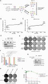
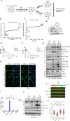
USP37 is a novel target showing synthetic lethality with BRCA1 loss. (A) Schematic of the CRISPR screen to define the genetic interaction with BRCA1 loss with or without treatment with 25 nM olaparib (Ola) in HeLa BRCA1 mAID cells. Ranking of co-essential genes in HeLa BRCA1−/− not treated with olaparib (B) or treated with olaparib (C) based on drugZ analysis of the results of CRISPR/Cas9 screen. The Z-score was used to define possible vulnerability to cell lethality with BRCA1 loss with or without treatment with olaparib. (D) Western blot analysis to confirm USP37 knockout (KO) in HeLa BRCA1 mAID cells. WT: wild type. (E) Representative crystal violet images of the clonogenic survival assay in USP37-depleted HeLa BRCA1 mAID cells treated with or without BRCA1 depletion and/or olaparib. (F) Quantification of the data in panel (E). Data are presented as means (±SD), n = 3 independent experiments, and P values were derived from a two-way ANOVA with Tukey’s multiple comparisons test. *P < .05, **P < .01, ***P < .001, ****P < .0001. (G) Western blot analysis to confirm USP37 KO in HEK293A BRCA1 dTAG cells with or without induction of BRCA1 degradation. (H) Representative crystal violet images of the clonogenic survival assay in USP37-depleted HEK293A BRCA1 dTAG cells treated with the indicated doses of dTAGV-1 (50 and 100 nM) or dTAGV-1 Neg (100 nM). (I) CellTiter-Glo assay to examine the synthetic lethality of USP37 loss and BRCA1 loss in HEK293A BRCA1 dTAG cells with various doses of dTAGV-1 NEG or dTAGV-1 treatment.
Figure 2.
Figure 2.
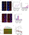
Depletion of USP37 results in genomic instability and uncontrolled degradation of nascent replication forks in BRCA1-deficient cells. (A) Representative images of γH2AX staining following treatment with olaparib (Ola; 2 μM) for 48 h in WT and USP37 KO HeLa BRCA1 mAID cells. BRCA1−/− cells were cells with BRCA1 degradation following treatment with 5-Ph-IAA and doxycycline. BRCA1+/+ were BRCA1 WT cells. NT: no treatment. Scale bar, 10 μm. (B) Statistical quantification of γH2AX foci formation per cell from panel (A). Mean number of γH2AX foci per cell was analyzed in >100 cells per sample. P values were derived from a one-way ANOVA with Tukey’s multiple comparisons test. ****P < .0001. (C) Statistical quantification of percentage of cells with micronuclei from panel (A). At least 100 cells were analyzed per sample over three independent experiments. Data are presented as means (±SD), n = 3 independent experiments, and P values were derived from a two-way ANOVA with Tukey’s multiple comparisons test. **P < .01. (D) Representative images of RAD51 staining following treatment with olaparib (2 μM) for 48 h in WT and USP37 KO HeLa BRCA1 mAID cells. Scale bar, 10 μm. (E) Quantification of data in panel (D). Data are presented as means (±SD), n = 3 independent experiments, and P values were derived from a two-way ANOVA with Tukey’s multiple comparisons test. ns: not significant. (F) Top, a schematic of IdU/CIdU pulse-labeling followed by a 4-h treatment with hydroxyurea (HU; 5 mM). Bottom, representative images of ldU and CIdU replication tracks in HU-treated WT or USP37 KO HeLa BRCA1+/+ and BRCA1−/− cells. (G) Dot plot of CIdU to ldU tract length ratios for individual replication forks in cells from panel (F). The median value of 100 or more IdU and CldU tracts per experimental condition is indicated. Data are presented as means (±SD), n = 3 independent experiments, and P values were derived from a one-way ANOVA with Tukey’s multiple comparisons test. *P < .05, **P < .01, ***P < .001, ****P < .0001.
Figure 3.
Figure 3.
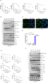
Loss of USP37 leads to vulnerability upon ATR or CHK1 inhibition (ATRi/CHK1i) or HU-induced replication stress. (A) Cell viability assays performed in HeLa wild-type (WT) and USP37 knockout (KO) cells. Cells were treated with the indicated doses of ATM inhibitor (ATMi, AZD0156), DNAPK inhibitor (DNAPKi, NU7441), ATRi (AZD6738), and CHK1i (LY2603618). Data are presented as mean ± SD. n = 3 independent biological experiments. (B) Western blot analysis of various DNA damage signals conducted in HeLa WT and USP37 KO cells upon treatment with 1 μM ATRi at different time points. (C) Representative images of RPA2 staining following treatment with ATRi (1 μM) for 48 h in HeLa WT and USP37 KO cells. NT: no treatment. Scale bar, 10 μm. (D) Quantification of percentage of cells from panel (C) with RPA2 foci >10. At least 100 cells were analyzed per sample over three biologically independent experiments. Data are presented as means (±SD), n = 3 independent experiments, and P values were derived from a two-way ANOVA with Tukey’s multiple comparisons test. ****P < .0001, ns: not significant. (E) Cell viability assays performed in HeLa WT and USP37 KO cells. Cells were treated with the indicated doses of IR, HU, camptothecin, mitomycin C, olaparib (Ola), or ultraviolet. Data are presented as mean ± SD. n = 2 independent biological experiments. (F) Western blot analysis conducted in HeLa WT and USP37 KO cells upon treatment with 2 mM HU at different time points to analyze various DNA damage signals with indicated antibodies.
Figure 4.
Figure 4.
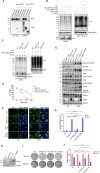
USP37 is required for cell survival in response to replication stress, and it may mediate RPA deubiquitination. (A) 293T cells were transfected with Myc-tagged USP37 and SFB-tagged RPA1, RPA2, or RPA3. Lysates were subjected to co-immunoprecipitation (IP) assays using streptavidin-binding beads and immunoblotted with indicated antibodies. (B) HEK293T cells were co-transfected with indicated plasmids for in vivo deubiquitination assay. After 48 h, cells were treated with MG132 for 6 h before harvest. SFB-RPA2 was immunoprecipitated by streptavidin beads. Blots were probed with the indicated antibodies. HU: hydroxyurea; WT: wild type. (C) HeLa WT and USP37 KO cells were co-transfected with indicated plasmids for in vivo deubiquitination assay. After 48 h, cells were treated with MG132 for 6 h before harvest. SFB-RPA2 was immunoprecipitated by streptavidin beads. Blots were probed with the indicated antibodies. (D) Cell viability assay in response to treatment with ATRi was performed in HeLa USP37 knockout (KO) cells reconstituted with USP37 WT and catalytic dead mutant C350S. Data are presented as means ± SD, n = 3 independent experiments. (E) Western blot analysis of various DNA damage signals conducted in HeLa USP37 KO cells reconstituted with USP37 WT and catalytic dead mutant C350S upon treatment with ATRi. NT: no treatment. (F) Representative images of RPA2 staining following treatment with ATRi (1 μM) for 48 h in HeLa USP37 KO cells reconstituted with USP37 WT and catalytic dead mutant C350S. Scale bar, 10 μm. (G) Quantification of percentage of cells from panel (F) with RPA2 foci >10. At least 100 cells were analyzed per sample over three biologically independent experiments. Data are presented as means (±SD), n = 3 independent experiments, and P values were derived from a two-way ANOVA with Tukey’s multiple comparisons test. ****P < .0001, ns: not significant. (H) Western blot analysis to evaluate the expression of USP37 WT and catalytic dead mutant C350S in HeLa BRCA1 mAID cells. (I) Representative crystal violet images of the clonogenic survival assay conducted in USP37-depleted HeLa BRCA1 mAID cells reconstituted with USP37 WT and catalytic dead mutant C350S. (J) Quantification of the data in panel (I). Data are presented as means (±SD), n = 3 independent experiments, and P values were derived from a two-way ANOVA with Tukey’s multiple comparisons test. **P < .01, ***P < .001, ns: not significant.
Figure 5.
Figure 5.
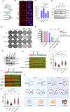
HLTF counteracts the effects of USP37 loss upon replication stress. (A) Schematic of the CRISPR screen to define the genetic interaction in HeLa WT and USP37 KO cells with or without treatment with ATRi (0.2 μM). Ranking of co-essential genes in HeLa USP37 KO cells treated with ATRi (B) or without ATRi (C) based on drugZ analysis of the results of CRISPR/Cas9 screen. The Z-score was used to define possible vulnerability to cell lethality with USP37 loss with or without treatment with ATRi. (D) Western blot analysis to confirm HLTF KO in HeLa WT and USP37 KO cells. (E) CellTiter-Glo assay to reveal synthetic lethality upon USP37 loss and HLTF loss in HeLa cells with various doses of ATRi. (F) CellTiter-Glo assay to reveal synthetic lethality upon USP37 loss and HLTF loss in HeLa cells with various doses of HU. (G) HeLa WT, USP37 KO, HLTF KO, and USP37&HLTF DKO cells were treated with 1 μM ATRi or 1 μM CHK1i for 24 h. Cell lysates were examined by western blot analysis to evaluate various DNA damage signals with indicated antibodies. NT: no treatment. (H) Representative images of RPA2 staining following treatment with ATRi (1 μM) for 48 h in HeLa WT, USP37 KO, HLTF KO, and USP37&HLTF DKO cells. Scale bar, 10 μm. (I) Quantification of percentage of cells from panel (H) with >10 RPA2 foci. At least 100 cells were analyzed per sample over three biologically independent experiments. Data are represented as means (±SD), n = 3 independent experiments, and P values were derived from a two-way ANOVA with Tukey’s multiple comparisons test. ****P < .0001, ns: not significant. (J) Top, schematic of IdU/CIdU pulse-labeling. Bottom, HeLa WT, USP37 KO, HLTF KO, and USP37&HLTF DKO cells were sequentially labeled in IdU for 30 min followed by CIdU in the presence of ATRi (0.5 μM) for 30 min. Representative images of ldU and CIdU replication tracks in ATRi-treated indicated cells are shown. (K) Dot plot of CIdU to ldU tract length ratios for individual replication forks in cells from panel (J). The median value of 100 or more IdU and CldU tracts per experimental condition is indicated. Data are presented as means (±SD), n = 3 independent experiments, and P values were derived from a one-way ANOVA with Tukey’s multiple comparisons test. **P < .01, ***P < .001, ****P < .0001. (L) HeLa WT, USP37 KO, HLTF KO, and USP37&HLTF DKO cells were treated with 1 μM CHK1i or 2 mM HU for 24 h. Chromatin fractions were harvested and examined by western blot analysis with indicated antibodies.
Figure 6.
Figure 6.
Depletion of HLTF rescues cell lethality upon USP37 loss in BRCA1-deficient cells, and it prevents MRE11-dependent nascent fork degradation. (A) Top: Schematic of the native BrdU assay. Bottom: Model for the native BrdU assay to detect the reversed replication fork. (B) Representative images of native BrdU staining in HeLa WT, USP37 KO, HLTF KO, and USP37&HLTF DKO cells with or without the treatment of HU (4 mM) for 3 h. Scale bar, 10 μm. (C) Quantification of percentage of cells from panel (B) with BrdU foci. At least 100 cells were analyzed per sample over three biologically independent experiments. Data are represented as means (±SD), n = 3 independent experiments, and P values were derived from a two-way ANOVA with Tukey’s multiple comparisons test. *P< .05, **P < .01, ***P < .001, ****P < .0001, ns: not significant. (D) Western blot analysis to confirm HLTF KO in HeLa BRCA1 mAID WT and USP37 KO cells with or without induction of BRCA1 degradation. (E) Representative crystal violet images of the clonogenic survival assay in cells from panel (D) with or without treatment with olaparib (Ola). (F) Quantification of the data in panel (E). Data are presented as means (±SD), n = 3 independent experiments, and P values were derived from a one-way ANOVA with Tukey’s multiple comparisons test. *P < .05, **P < .01. (G) Western blot analysis to check siRNA knockdown efficiency of USP37 and HLTF in SUM149PT cells. (H) Top: Schematic of IdU/CIdU pulse-labeling followed by a 4-h treatment with HU (2 mM). Bottom, representative images of ldU and CIdU replication tracks in HU-treated indicated cells. (I) Dot plot of CIdU to ldU tract length ratios for individual replication forks in cells from panel (H). The median value of 100 or more IdU and CldU tracts per experimental condition is indicated. Data are presented as means (±SD), n = 3 independent experiments, and P values were derived from a one-way ANOVA with Tukey’s multiple comparisons test. ****P < .0001. (J) Top: Schematic of IdU/CIdU pulse-labeling followed by a 4-h treatment with or without Mirin (50 μM) together with HU (2 mM). Bottom, representative images of ldU and CIdU replication tracks in indicated treated cells. (K) Dot plot of CIdU to ldU tract length ratios for individual replication forks in cells from (J). The median value of 100 or more IdU and CldU tracts per experimental condition is indicated. Data are presented as means (±SD), n = 3 independent experiments, P values were derived from a one-way ANOVA with Tukey’s multiple comparisons test. ****P < .0001. (L) Model of the mechanism of synthetic lethality in BRCA1-deficiency and USP37 loss.

References

    1. Petrucelli N, Daly MB, Feldman GL Hereditary breast and ovarian cancer due to mutations in BRCA1 and BRCA2. Nature. 2010; 12:382–7. 10.1097/GIM.0b013e3181d38f2f. - DOI - PubMed
    1. Roy R, Chun J, Powell SN BRCA1 and BRCA2: different roles in a common pathway of genome protection. Nat Rev Cancer. 2011; 12:68–78. 10.1038/nrc3181. - DOI - PMC - PubMed
    1. Kass EM, Jasin M Collaboration and competition between DNA double-strand break repair pathways. FEBS Lett. 2010; 584:3703–8. 10.1016/j.febslet.2010.07.057. - DOI - PMC - PubMed
    1. Hashimoto Y, Ray Chaudhuri A, Lopes M et al. Rad51 protects nascent DNA from Mre11-dependent degradation and promotes continuous DNA synthesis. Nat Struct Mol Biol. 2010; 17:1305–11. 10.1038/nsmb.1927. - DOI - PMC - PubMed
    1. Lomonosov M, Anand S, Sangrithi M et al. Stabilization of stalled DNA replication forks by the BRCA2 breast cancer susceptibility protein. Genes Dev. 2003; 17:3017–22. 10.1101/gad.279003. - DOI - PMC - PubMed

MeSH terms

LinkOut - more resources