. 2025 Jun 23;82(1):246.
doi: 10.1007/s00018-025-05760-3.

THAP11-mediated K48- and K63-linked ubiquitination is essential for the degradation of porcine reproductive and respiratory syndrome virus nonstructural protein 1β

Affiliations

THAP11-mediated K48- and K63-linked ubiquitination is essential for the degradation of porcine reproductive and respiratory syndrome virus nonstructural protein 1β

Binghua Chen et al. Cell Mol Life Sci. .

Abstract

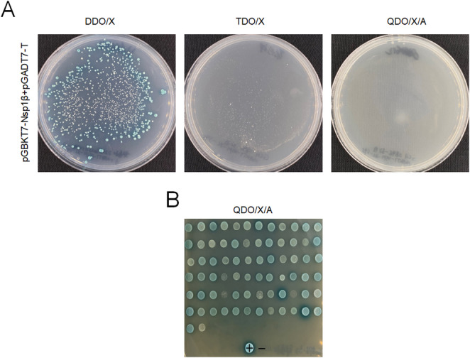
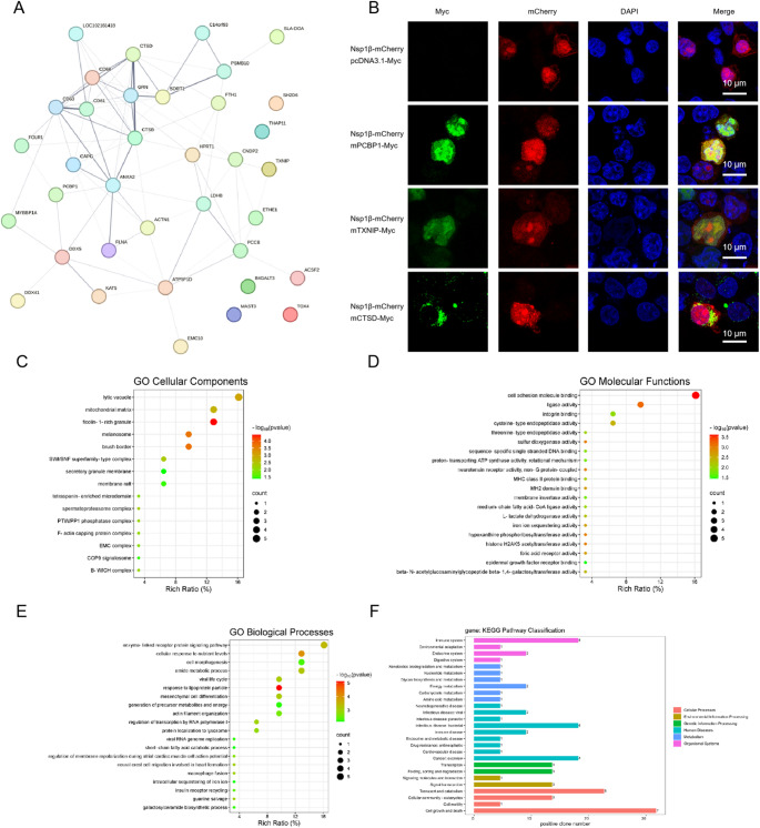
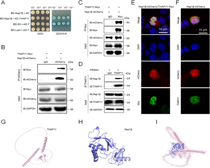
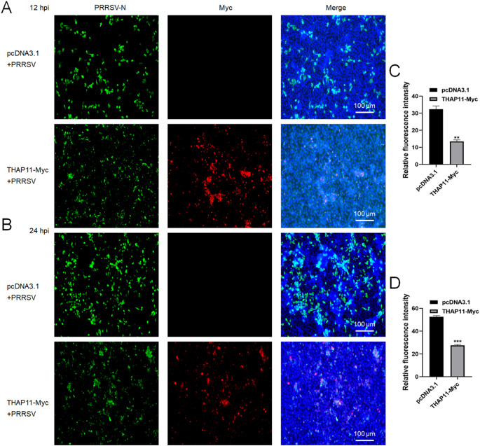
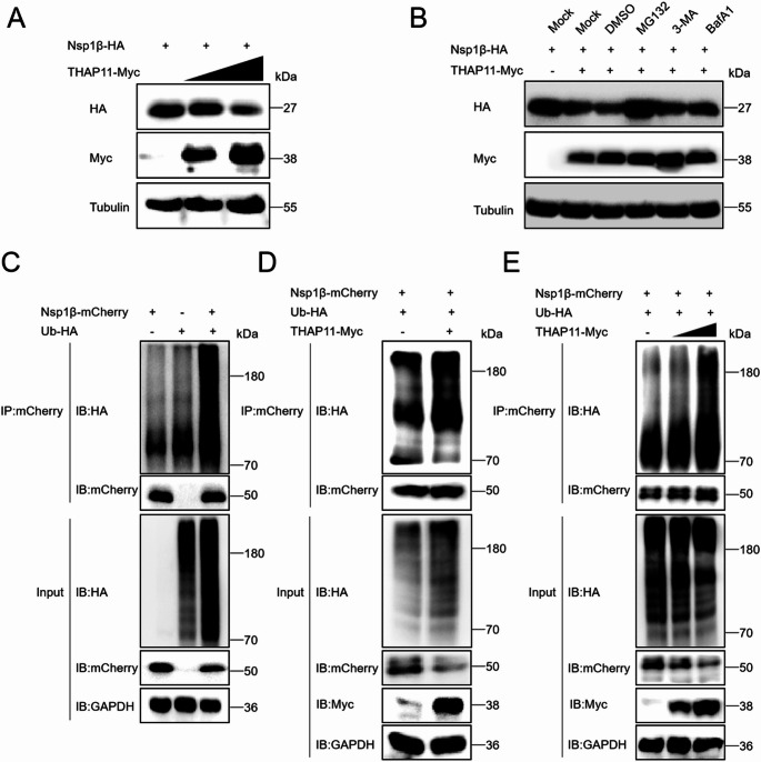
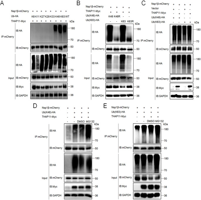
Porcine reproductive and respiratory syndrome virus (PRRSV) is a highly infectious pathogen in the global pig industry that causes significant economic losses. Owing to its rapid mutation, effective antiviral treatments or vaccines are still lacking. Therefore, it is essential to identify potential host factors that interact with PRRSV-encoded proteins. In this study, a porcine alveolar macrophage cDNA library was used to identify host proteins that interact with PRRSV nonstructural protein 1β (Nsp1β) via a yeast two-hybrid system. A total of 34 potential host factors were identified, with Thanatos-associated protein 11 (THAP11) strongly interacting with Nsp1β. These interactions were further analyzed via Gene Ontology (GO) and Kyoto Encyclopedia of Genes and Genomes (KEGG) pathway analyses. Co-localization of Nsp1β with THAP11, poly(rC)-binding protein 1 (PCBP1), thioredoxin-interacting protein (TXNIP), and cathepsin D (CTSD) was observed, and co-IP assays confirmed the Nsp1β-THAP11 interaction. The overexpression of THAP11 reduced PRRSV N protein accumulation, indicating an antiviral effect, whereas the silencing of THAP11 increased PRRSV replication. Furthermore, THAP11 promoted the degradation of Nsp1β by increasing K48- and K63-linked ubiquitination, thereby restricting PRRSV replication. These findings suggest that THAP11 exerts an antiviral effect by interacting with and degrading Nsp1β via the ubiquitin-proteasome system, providing insights for future PRRSV defence strategies.

Keywords: Nsp1β; PRRSV; THAP11; Virus-host interactions; Yeast two-hybrid screening.


Conflict of interest statement

Declarations. Competing interest: The authors declare that they have no conflicts of interest.

Figures

Fig. 1
Fig. 1
Detection of self-activation of the PRRSV Nsp1β protein via Y2H screening assays. (A) The pGBKT7-Nsp1β and pGADT7-T plasmids were cotransformed into Y2H Gold cells, which were then plated onto deficient culture media lacking Leu and Trp (DDO/X); Leu, Trp, and His (TDO/X); and Leu, Trp, His, and Ade (QDO/X/A), supplemented with X-α-gal and AbA. (B) Potential positive clones that interact with Nsp1β were screened on QDO/X/A media. The blue colonies indicate positive interactions, with “+” representing a positive result and “−” indicating a negative result
Fig. 2
Fig. 2
Bioinformatics analysis and colocalization of interacting proteins. (A) Candidate host protein interaction network. (B) Confocal fluorescence microscopy confirmed the co-localization of Nsp1β with the host proteins PCBP1, TXNIP, and CTSD. HEK293 T cells were cotransfected with mPCBP1-Myc, mTXNIP-Myc, or mCTSD-Myc along with Nsp1β-mCherry plasmids. Myc empty vector and Nsp1β-mCherry were used as controls. At 24 hpt, the cells were subjected to indirect immunofluorescence analysis and visualized via confocal microscopy. Scale bars, indicated by white lines, represent the magnification. The “m” notation indicates that the species is a monkey. (C) GO cellular components analysis of the interacting proteins from Nsp1β bait screening, revealing the cellular locations where these proteins are likely to function. (D) GO molecular functions analysis of proteins interacting with Nsp1β, as identified via bait screening. This analysis highlights the molecular functions associated with the interaction partners. (E) GO biological processes analysis of the proteins interacting with Nsp1β, providing insights into the biological processes in which these proteins are involved. (F) KEGG pathway analysis of the interacting proteins from Nsp1β bait screening, outlining the key signaling pathways and processes in which these proteins may participate
Fig. 3
Fig. 3
Nsp1β interacts with THAP11. (A) Y2H assays confirmed the interaction between Nsp1β and THAP11. The pGBKT7-Nsp1β and pGADT7-THAP11 plasmids, or an empty vector, were cotransformed into Y2H Gold cells. pGBKT7-53 + pGADT7-T served as a positive control, and pGBKT7-Lam + pGADT7-T was used as a negative control. The transformed cells were grown on DDO and QDO/X/A media, with serial dilutions applied to assess the interaction. (B) Co-IP assays confirmed that Nsp1β interacts with THAP11. Anti-mCherry or anti-IgG antibodies were conjugated to magnetic A beads, and Myc-tagged proteins were detected in the immunoprecipitate. (C) Co-IP using anti-Myc antibodies conjugated to magnetic A beads detected the Nsp1β-mCherry protein in the immunoprecipitate, further supporting the interaction between Nsp1β and THAP11. (D) Co-IP assays confirmed the interaction between endogenous THAP11 and Nsp1β in PRRSV-infected Marc-145 cells. Anti-THAP11 antibodies linked to magnetic A beads were used to detect Nsp1β in the immunoprecipitate. (E) Cotransfection of Nsp1β-mCherry and THAP11-Myc into HEK293 T cells for 24 h resulted in co-localization of the two proteins, as observed by confocal fluorescence microscopy. (F) Transfection of Nsp1β-mCherry into HEK293 T cells for 24 h showed colocalization of the Nsp1β and endogenous THAP11, as observed by confocal fluorescence microscopy. (G) The predicted spatial structure of the monkey THAP11 protein, as determined by the AlphaFold3 server, reveals its folding pattern and key structural features. (H) The predicted spatial structure of Nsp1β, also obtained through AlphaFold3, shows the structural arrangement of this viral protein involved in immune modulation. (I) The predicted binding interface between Nsp1β and THAP11, as modeled by AlphaFold3, illustrates the potential interaction site, highlighting how these two proteins might interact at the molecular level. Pink stands for THAP11 protein spatial folding layers, and blue stands for Nsp1β protein spatial folding fragments
Fig. 4
Fig. 4
THAP11 negatively regulates PRRSV infection. (A) Western blot analysis revealed endogenous THAP11 protein levels at different time points (0, 6, 12, 24, and 36 h) following PRRSV infection, revealing that THAP11 protein accumulation remained relatively stable throughout the infection. (B & C) Marc-145 cells were transfected with either 2 µg of THAP11-Myc or the pcDNA3.1 plasmid as a control. At 12 hpt, the cells were infected with PRRSV at an MOI of 0.5 for 12, 24, or 36 h. The accumulation of the PRRSV N protein was assessed via western blotting and relative expression of PRRSV ORF7 mRNA was analyzed by qPCR, which revealed reduced PRRSV replication in THAP11-Myc-expressing cells than in control cells. (D & E) Transfecting THAP11-Myc or empty vector plasmid into Marc-145 cells, the virus supernatant of 36 hpi was collected for plaque assay and virus titer analysis. (F & G) Knockdown of THAP11 enhanced PRRSV replication. Two siRNAs targeting THAP11 or a negative control siRNA were transfected into Marc-145 cells cultured in 12-well plates. At 12 hpt, the cells were infected with PRRSV for 36 h. The mRNA expression levels of THAP11 and the PRRSV ORF7 gene were measured via qPCR, and protein accumulation was analyzed via western blotting
Fig. 5
Fig. 5
IFA assays confirmed that THAP11 suppresses PRRSV replication. (A & B) Marc-145 cells were transfected with either THAP11-Myc or the pcDNA3.1 empty vector for 12 h, followed by PRRSV infection for 12–24 h. After the infection period, the cells were incubated overnight with the primary antibodies anti-N and anti-Myc. An anti-mouse 488 fluorescent secondary antibody was used to label the N protein, while an anti-rabbit 555 fluorescent secondary antibody was used to detect THAP11. Fluorescence images were captured via a Leica fluorescence microscope (Nikon, Germany), which revealed that THAP11 expression reduced PRRSV replication, as indicated by decreased N protein fluorescence. (C & D) Fluorescence count and luminance were statistically analyzed by Image J at 12 and 24 hpi
Fig. 6
Fig. 6
THAP11 promotes Nsp1β ubiquitination and degradation via the ubiquitin-proteasome pathway. (A) HEK293 T cells were cotransfected with Nsp1β-HA and various doses (0, 1.0, or 1.5 µg) of THAP11-Myc plasmids. At 24 hpt, the accumulation of the Nsp1β protein was analyzed via western blotting, which revealed that the Nsp1β protein was regulated in a dose-dependent manner by THAP11. (B) A total of 2.0 µg of Nsp1β-HA and THAP11-Myc or empty vector was cotransfected into HEK293 T cells for 24 h. At 10 hpt, the cells were treated with the proteasome inhibitor MG132, the autophagy inhibitors 3-MA and BafA1, or DMSO as a control. Western blot analysis detected the accumulation of Nsp1β after 24 h. (C) HEK293 T cells were cotransfected with Nsp1β-mCherry and Ub-HA plasmids or with Nsp1β-mCherry or Ub-HA plasmids alone as controls. At 24 hpt, the degree of Nsp1β ubiquitination was detected via co-IP assays with an anti-HA antibody. An anti-mCherry antibody was linked to magnetic A beads for immunoprecipitation, confirming the ubiquitination of Nsp1β. (D) HEK293 T cells were cotransfected with Nsp1β-mCherry, Ub-HA, and THAP11-Myc or pcDNA3.1 plasmids. At 24 hpt, western blot analysis revealed increased ubiquitination of Nsp1β in the presence of THAP11, confirming its role in promoting Nsp1β ubiquitination. (E) HEK293 T cells were cotransfected with Nsp1β-mCherry, Ub-HA, or various doses of the THAP11-Myc plasmid. At 24 hpt, western blot analysis was used to detect the ubiquitination levels of Nsp1β
Fig. 7
Fig. 7
THAP11-mediated Nsp1β degradation is dependent on K48- and K63-linked ubiquitination. (A) HEK293 T cells were co-transfected with Nsp1β-mCherry, THAP11-Myc, and ubiquitin mutants (K6, K11, K27, K29, K33, K48 or K63) or wild ubiquitin plasmids. At 24 hpt, western blot analysis detected Nsp1β ubiquitination levels, anti-mCherry was used in Co-IP assays. (B) HEK293 T cells were cotransfected with Nsp1β-mCherry, THAP11-Myc, or mutant Ub (K48, K48R, K63, or K63R) plasmids. At 24 hpt, western blot analysis was used to detect Nsp1β ubiquitination levels, and anti-mCherry was used in Co-IP assays. (C) HEK293 T cells were cotransfected with Nsp1β-mCherry, Ub(K48)-HA and THAP11-Myc or pcDNA3.1 plasmids or cotransfected with Nsp1β-mCherry, Ub(K63)-HA and THAP11-Myc or pcDNA3.1 plasmids. At 24 hpt, the degree of Nsp1β ubiquitination was detected via co-IP assays with an anti-HA antibody. mCherry antibody was coupled to magnetic A beads for immunoprecipitation. (D & E) HEK293 T cells were co-transfected with Nsp1β-mCherry, THAP11-Myc (empty vector control), and ubiquitin mutants (K48 or K63) plasmids. At 10 hpt, cells were treated with MG132 (10 µM) or DMSO for 14 h, western blot analysis detected Nsp1β ubiquitination levels, and anti-mCherry was used in Co-IP assays

Similar articles

Cited by

References

    1. Lunney JK, Fang Y, Ladinig A, Chen N, Li Y, Rowland B, Renukaradhya GJ (2016) Porcine reproductive and respiratory syndrome virus (PRRSV): pathogenesis and interaction with the immune system. Annu Rev Anim Biosci 4:129–154 - PubMed
    1. Kwon B, Ansari IH, Pattnaik AK, Osorio FA (2008) Identification of virulence determinants of Porcine reproductive and respiratory syndrome virus through construction of chimeric clones. Virology 380:371–378 - PubMed
    1. Li Y, Wang X, Jiang P, Chen W, Wang X (2008) Genetic analysis of two Porcine reproductive and respiratory syndrome viruses with different virulence isolated in China. Arch Virol 153:1877–1884 - PubMed
    1. Darwich L, Gimeno M, Sibila M, Diaz I, de la Torre E, Dotti S, Kuzemtseva L, Martin M, Pujols J, Mateu E (2011) Genetic and Immunobiological diversities of Porcine reproductive and respiratory syndrome genotype I strains. Vet Microbiol 150:49–62 - PubMed
    1. Liu J, Wei C, Lin Z, Fan J, Xia W, Dai A, Yang X (2019) Recombination in lineage 1, 3, 5 and 8 of Porcine reproductive and respiratory syndrome viruses in China. Infect Genet Evol 68:119–126 - PubMed

MeSH terms

Substances

LinkOut - more resources