

doi: 10.1007/s00401-025-02907-1.
CNS methylation classifiers may misclassify normal developing cerebellar cortex as medulloblastoma
Affiliations
- PMID: 40549014
- PMCID: PMC12185629
- DOI: 10.1007/s00401-025-02907-1
Item in Clipboard
CNS methylation classifiers may misclassify normal developing cerebellar cortex as medulloblastoma
Acta Neuropathol.
.
No abstract available
Conflict of interest statement
Declarations. Conflict of interest: The authors declare no competing interests.
Figures
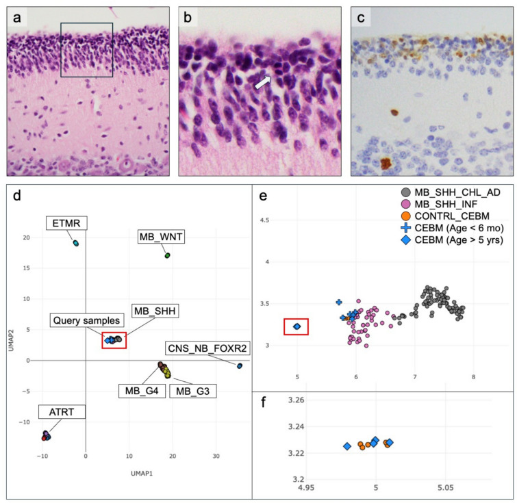
Appearance of cerebellar external granule cell layer and performance of methylation classification on cerebellar cortex samples before 6 months of age. In late gestation and infancy, the external granular layer (EGL) covers the cerebellar cortex, as seen in CHLA_CB_2 (a). The EGL consists of closely packed primitive-appearing cells with nuclear angulation (b, enlarged from boxed area shown in a) and, at times, irregular contours. Apoptotic bodies and mitotic figures (b, arrow) are common in the EGL. Ki-67 labeling index is elevated in the EGL due to ongoing cell proliferation (c). These features are all developmentally appropriate but can be misinterpreted as subpial spread of an embryonal neoplasm. Using DNA methylation array data, UMAP visualization (d–f) shows immature cerebellar tissue samples (blue crosses) more closely associate with MB_SHH groups (e, enlarged from boxed area shown in (d). Older pediatric samples (blue diamonds) show clustering away from those representing individuals aged < 6 months, and associate with reference controls (f, enlarged from boxed area shown in e)
References
LinkOut - more resources
Full Text Sources
