

A Comprehensive Review of Kimura Disease
- PMID: 40549072
- PMCID: PMC12185846
- DOI: 10.1007/s12105-025-01812-z
A Comprehensive Review of Kimura Disease
Abstract
Purpose: Kimura disease (KD) is a rare, chronic inflammatory disorder that primarily affects the head and neck regions, often mimicking neoplastic conditions. This study aims to provide a comprehensive review of KD, focusing on its clinical presentation, diagnostic challenges, optimal management strategies, and primary histopathologic differential diagnosis.
Methods: A systematic review of literature was conducted using PubMed, Scopus, and Google Scholar databases. We analyzed case reports, retrospective studies, and clinical trials published in English. We extracted data on epidemiology, clinical presentation, laboratory findings, histologic features, current understanding of the pathogenesis, treatment, and prognosis.
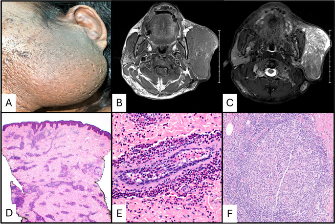
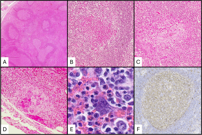
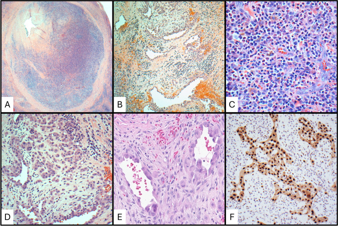
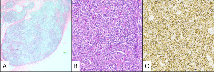
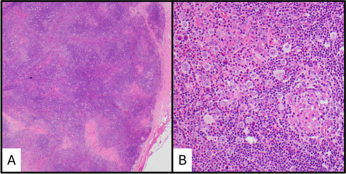
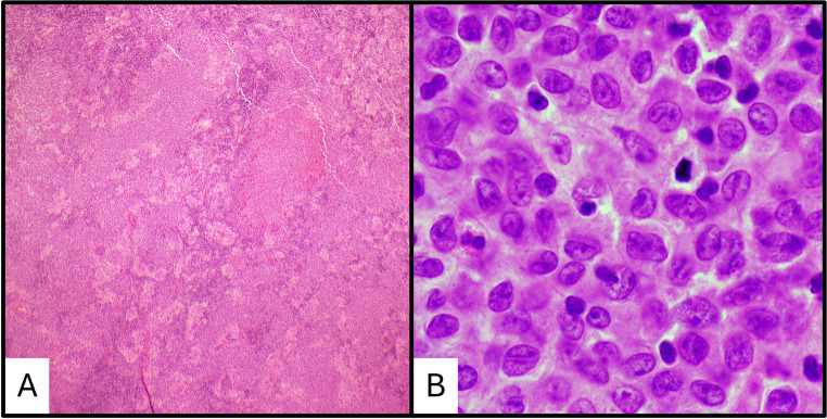
Results: KD predominantly affects young Asian males, presenting with painless subcutaneous masses, peripheral eosinophilia, and elevated serum IgE levels. Histopathology reveals lymphoid follicular hyperplasia with eosinophilic infiltration. Biopsy is required for diagnosis. The pathogenesis of KD is poorly understood, but recent studies have elucidated some potentially important mechanisms of the disease. Treatment options include systemic corticosteroids, surgical excision, radiotherapy, and cytotoxic therapies, with recurrence rates varying among modalities.
Conclusion: KD remains a diagnostic challenge due to its overlapping features with a variety of neoplastic and non-neoplastic conditions. While corticosteroids offer temporary relief and can be useful in cases with renal involvement, surgical excision remains the most definitive treatment. Future research should focus on targeted therapies to improve long-term disease control and reduce recurrence.
Keywords: Eosinophilia; Head and neck pathology; Hematopathology; Kimura disease.
Conflict of interest statement
Declarations. Ethics Approval: This article does not contain any studies with human participants are animals performed by any of the authors. Consent to Participate: For this type of study informed consent is not required. Consent for Publication: For this type of study consent for publication is not required. Statistical Declaration: No statistical analysis was performed for this manuscript. Disclaimer: The views expressed herein are those of the authors and do not necessarily reflect the official policy or position of Walter Reed National Military Medical Center, Fort Belvoir Community Hospital, the U.S. Army Medical Department, the U.S. Army Office of the Surgeon General, the Department of the Air Force, the Department of the Army, Department of Defense, the Uniformed Services University of the Health Sciences, or any other agency of the U.S. Government. The identification of specific products or scientific instrumentation is considered an integral part of the scientific endeavor and does not constitute endorsement or implied endorsement on the part of the authors, DoD, or any component agency. Competing Interests: The authors declare no competing interests.
Figures
References
-
- Kim H Eosinophilic hyperplastic lymphogranuloma, comparison with mikulicz’s disease. Chi Med J 1937:699–670
-
- Kimura T On the unusual granulations combined with hyperplastic changes of lymphatic tissue. Trans Soc Pathol Jpn 1948:179–180
-
- Chen H, Thompson LDR, Aguilera NSI, Abbondanzo SL (2004) Kimura disease: a clinicopathologic study of 21 cases. Am J Surg Pathol 28:505–513. 10.1097/00000478-200404000-00010 - PubMed
-
- Wells GC, Whimster IW (1969) Subcutaneous angiolymphoid hyperplasia with eosinophilia. Br J Dermatol 81:1–14. 10.1111/j.1365-2133.1969.tb15914.x - PubMed
Publication types
LinkOut - more resources
Full Text Sources
