. 2025 Jul;122(26):e2426204122.
doi: 10.1073/pnas.2426204122. Epub 2025 Jun 23.

SARS-CoV-2 nucleocapsid protein directly prevents cGAS-DNA recognition through competitive binding

Affiliations

SARS-CoV-2 nucleocapsid protein directly prevents cGAS-DNA recognition through competitive binding

Theresia Gutmann et al. Proc Natl Acad Sci U S A. 2025 Jul.

Abstract

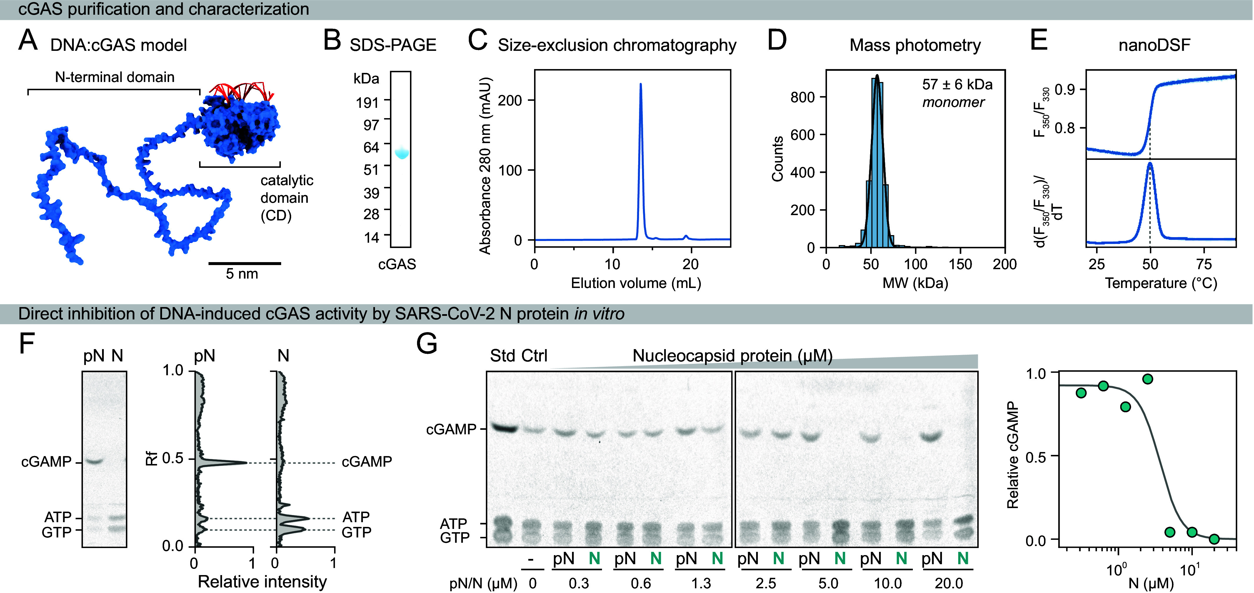
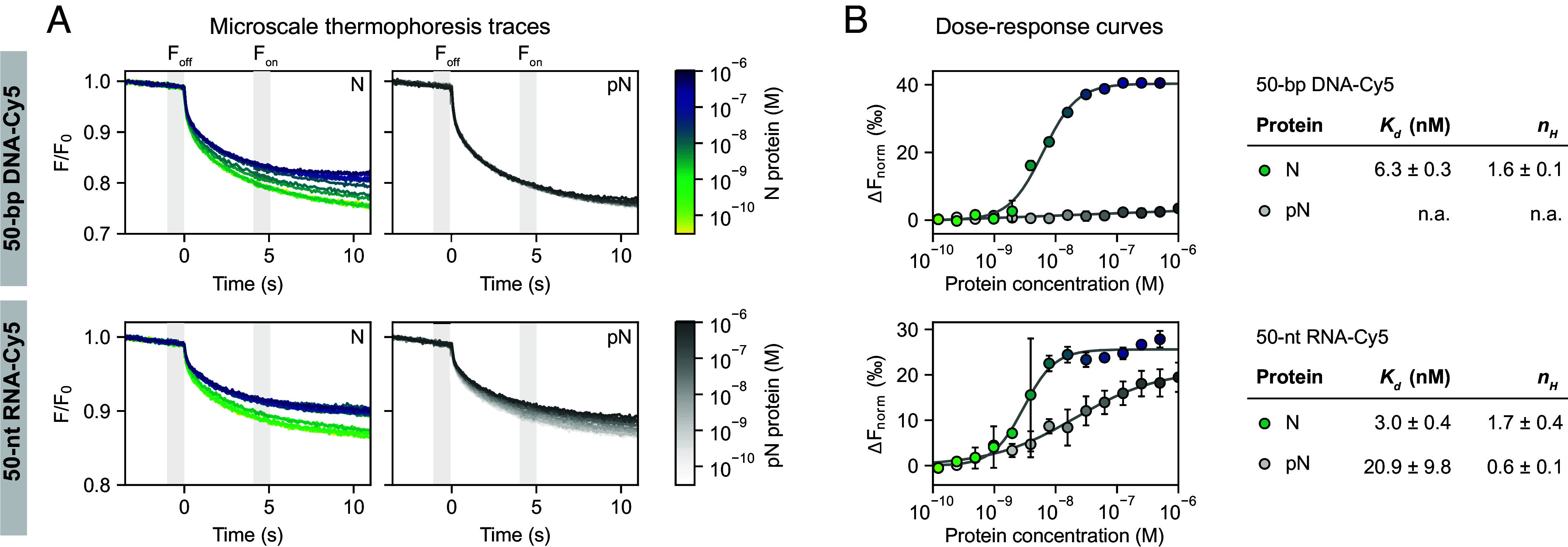
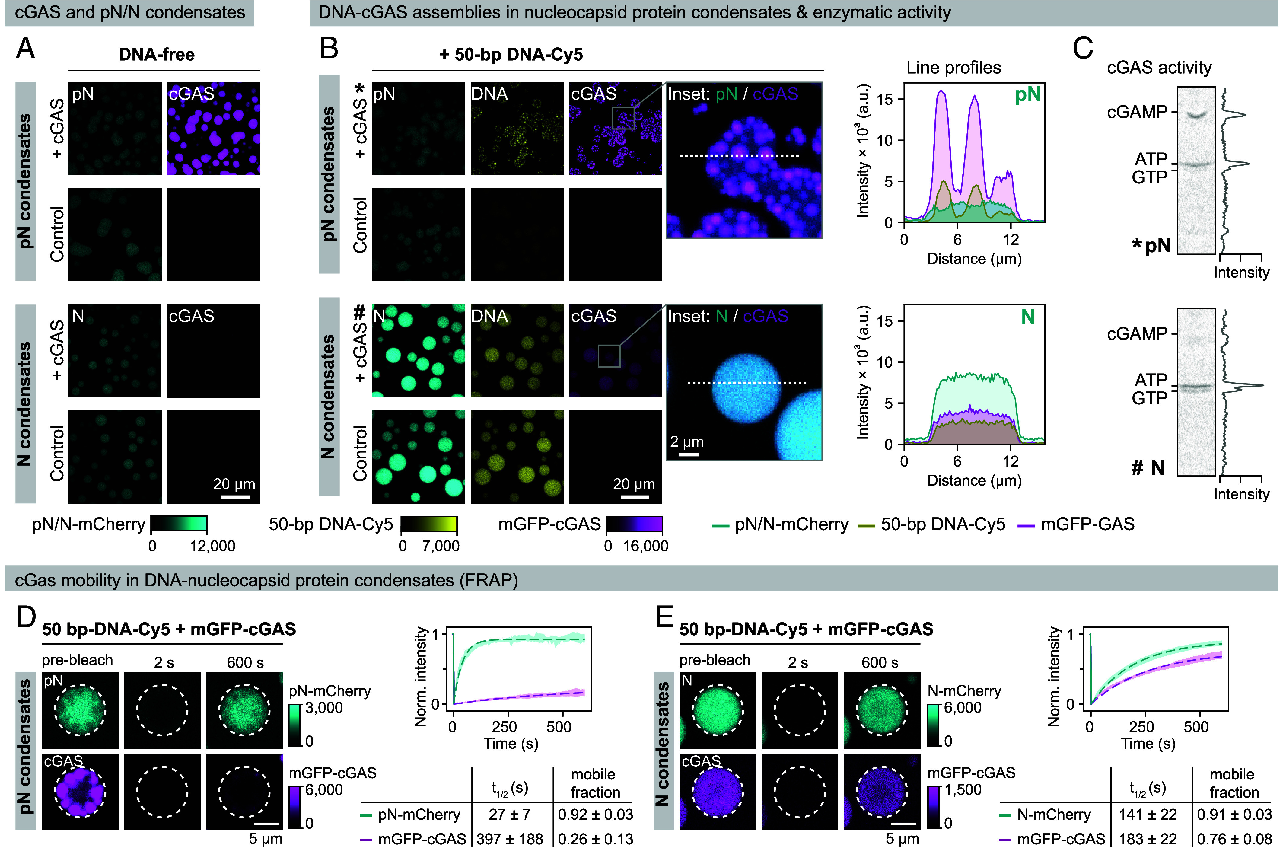
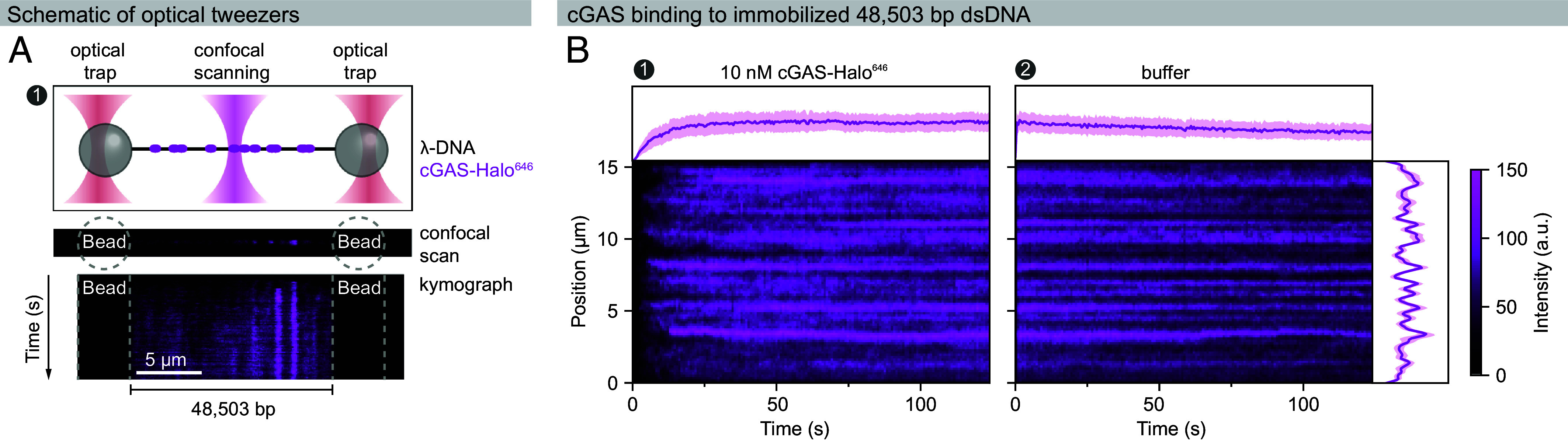
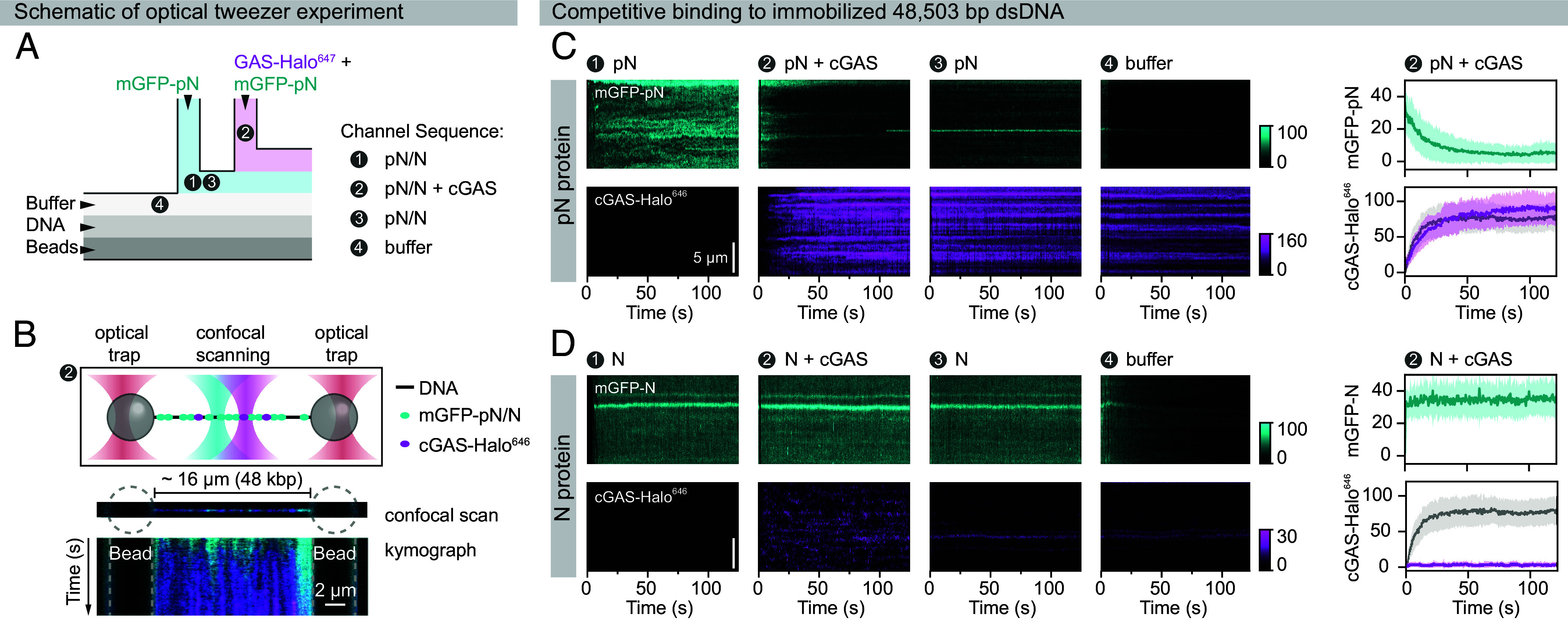
A hallmark of severe acute respiratory syndrome coronavirus 2 (SARS-CoV-2) infection is the delayed interferon response. Interferons are typically produced upon host recognition of pathogen- or damage-associated molecular patterns, such as nucleic acids. While the mechanisms by which SARS-CoV-2 evades host recognition of its RNA are well studied, how it evades immune responses to cytosolic DNA-leaked from mitochondria or nuclei during infection-remains poorly understood. Here, we demonstrate that the SARS-CoV-2 nucleocapsid protein directly suppresses DNA sensing by cyclic guanosine monophosphate-adenosine monophosphate synthase (cGAS). Although primarily known for packaging the viral RNA genome, we uncover that the SARS-CoV-2 nucleocapsid protein also binds DNA with high affinity and competitively blocks cGAS activation. Using cell-free biochemical and biophysical approaches, including single-molecule optical tweezers, we show that the nucleocapsid protein binds to DNA at nanomolar concentrations and cocondenses with DNA at micromolar concentrations, thereby impeding stable cGAS-DNA interactions required for signal propagation. Hyperphosphorylation of the nucleocapsid protein diminishes its competitive binding capacity. Our findings reveal an unexpected role of the SARS-CoV-2 nucleocapsid protein in directly suppressing the cGAS-STING pathway, strongly suggesting that this contributes to the delayed interferon response during infection. This study raises the possibility that nucleocapsid proteins of other RNA viruses may also exhibit moonlighting functions by antagonizing host nucleic acid-sensing pathways.

Keywords: biomolecular condensates; cGAS; coronavirus; innate immune system; nucleocapsid protein.


Conflict of interest statement

Competing interests statement:A.A.H. is a founder of and consultant for Dewpoint Therapeutics Inc. The company was not involved in the work of this manuscript.

Figures

Fig. 1.
Fig. 1.
Characterization of recombinant cGAS and inhibition of its activity by the SARS-CoV-2 nucleocapsid protein. (A) Structural model of full-length human cGAS (blue) bound to 17-bp DNA (red) at the primary DNA-binding site, based on the crystal structure of the catalytic domain [PDB: 6CT9, (34)]. The disordered N-terminal domain was modeled using AlphaFold, with torsion angles adjusted for optimal visualization. (B) Coomassie-stained SDS-PAGE gel of purified human cGAS. (C) Size-exclusion chromatography confirms the homogeneity of recombinant cGAS, which elutes as a monomer from a Superdex 200 Increase 10/300 GL column. (D) Mass photometry analysis indicated a molecular weight of 59 ± 7.5 kDa, consistent with monomeric full-length cGAS. (E) The thermal stability of cGAS was measured by nanoDSF, which assessed the intrinsic fluorescence ratio at 350 and 330 nm (n = 2; error bands represent SD). A dashed line indicates the melting temperature of 49.80 ± 0.04 °C. (F) In vitro synthesis of 2′3′-cGAMP by cGAS in the presence of 0.2 µM 100-bp DNA and 10 µM unphosphorylated (N) or phosphorylated (pN) nucleocapsid protein in 120 mM NaCl, 20 mM HEPES, pH 7.5, 5 mM MgCl2, 0.5 mM ATP, 0.5 mM GTP, and 1 mM DTT. The thin-layer chromatography plate shows separated nucleotides (Left) with the corresponding densitograms (Right). (G) Dose-dependent inhibition of cGAS (1 µM) activity in the presence of 0.2 µM 100-bp DNA and increasing concentrations of N or pN protein. The Right panel shows cGAMP production relative to the nucleocapsid protein-free control.
Fig. 2.
Fig. 2.
SARS-CoV-2 N protein binds to DNA and RNA with high affinity. Microscale thermophoresis was used to characterize nucleic acid binding by nucleocapsid proteins in a buffer containing 20 mM HEPES, pH 7.5, 150 mM NaCl, 0.05% Tween-20, and 1 mM DTT. (A) Normalized microscale thermophoresis traces show the binding of label-free N and pN proteins to 0.75 nM of Cy5-labeled 50-bp DNA (Top) and 50-nt RNA (Bottom). Gray-shaded time intervals were analyzed to generate dose–response curves shown in (B). Each trace is color-coded by protein concentration. (B) Corresponding dose–response curves for N and pN protein binding to DNA (Top) and RNA (Bottom) were generated by fitting the change in thermophoretic mobility to the Hill equation. Points represent the mean of two independent experiments, color-coded as in (A), with error bars representing SD. Apparent dissociation constants (Kd) and Hill coefficients (nH) are reported ± SD.
Fig. 3.
Fig. 3.
DNA–cGAS clusters do not form within nucleocapsid protein condensates. Condensates were formed by mixing pN or N protein (10 µM, including 10 mol% of the respective mCherry-labeled nucleocapsid) in a buffer containing 50 mM NaCl, 20 mM HEPES, pH 7.5, 5 mM MgCl2, 0.1 mM ATP, 0.1 mM GTP, 5% glycerol, 2 mM DTT; in the presence or absence of mGFP-cGAS (1 µM) and/or 50-bp DNA-Cy5 (0.5 µM). (A) Confocal microscopy images showing partitioning of mGFP-cGAS into DNA-free pN (Top) and N protein (Bottom) condensates. mGFP-cGAS partitions preferentially into pN protein condensates. (B) Confocal microscopy images showing partitioning of mGFP-cGAS into DNA-containing pN (Top) and N protein (Bottom) condensates. DNA has a high affinity for the unphosphorylated N protein and enhances N protein condensation and protein density in the condensed phase. Magnified merged images on the right demonstrate the DNA–cGAS clustering in pN versus homogeneous partitioning in N protein condensates. Fluorescence signal intensities along the dashed lines are shown in the graphs. (C) cGAS activity in the presence of DNA and pN (*) or N protein condensates (#) monitored by thin-layer chromatography. (D and E) FRAP analysis reveals differential mGFP-cGAS mobility in the presence of DNA and pN (D) or N protein condensates (E). Panels show representative snapshots before, at 2 s, and at 600 s after bleaching. Fluorescence intensities were fitted to a diffusion model. Graphs show the fitted curves (dashed line), and the error band represents the mean intensities ± SD (n = 7 droplets). mGFP-cGAS forms immobile clusters in the presence of pN protein condensates and retains mobility within N-DNA condensates.
Fig. 4.
Fig. 4.
Real-time visualization of cGAS binding to long DNA using single-molecule optical tweezers. The dynamic binding and unbinding of cGAS-Halo646 to a single DNA molecule were monitored using optical tweezers. Experiments were conducted in a buffer containing 50 mM NaCl, 20 mM HEPES, pH 7.0, 5% glycerol, 2 mM DTT, and 0.3 mg/mL BSA. (A) Schematic presenting the optical tweezer setup used to study cGAS-DNA interactions. Biotinylated λ-phage DNA (black) is tethered between two optically trapped, streptavidin-functionalized polystyrene beads. The DNA is exposed to cGAS-Halo646, which binds and forms clusters along the DNA (magenta). Confocal microscopy is used to scan the DNA tether. Kymograph projections along the DNA visualize the spatiotemporal dynamics of protein-DNA interactions. (B) Representative kymographs show cGAS-Halo646 interactions with DNA at 10 nM (Left) and after transferring the DNA tether to buffer (Right). The corresponding fluorescence intensity profiles are plotted over time (above kymographs) or along the DNA (Left), representing mean pixel intensity ± SD. cGAS-Halo646 binds rapidly to DNA and forms stable clusters that persist after the DNA tether is transferred to plain buffer, reflecting the multivalent interactions and resulting high avidity within cGAS-DNA clusters.
Fig. 5.
Fig. 5.
Competitive binding of SARS-CoV-2 nucleocapsid proteins to long DNA revealed by single-molecule optical tweezer assays. Simultaneous binding and unbinding of mGFP-labeled SARS-CoV-2 nucleocapsid proteins and cGAS-Halo646 to a single DNA molecule were monitored in real time using optical tweezers. Experiments were performed in a buffer containing 50 mM NaCl, 20 mM HEPES, pH 7.0, 5% glycerol, 2 mM DTT, and 0.3 mg/mL BSA. (A and B) Schematic of the optical tweezer microfluidic setup. Biotinylated λ-phage DNA was tethered between two optically trapped streptavidin-functionalized beads, then sequentially moved through four channels: (step ➊) 100 nM mGFP-N or mGFP-pN; (step ➋) 100 nM nucleocapsid protein and 10 nM cGAS–Halo646; (step ➌) nucleocapsid protein alone (100 nM); and (step ❹) plain buffer. Confocal line scans were used to generate kymographs across the DNA (48,503 bp) to visualize the DNA-binding kinetics of the proteins. (C and D) Representative kymographs showing mGFP-pN (C) or mGFP-N (D) binding to immobilized DNA and competition with cGAS–Halo646. For step ➋, pixel intensities are plotted as mean ± SD. Gray traces indicate cGAS–Halo646 intensities in the absence of nucleocapsid proteins (from Fig. 4B). While cGAS-Halo646 induces rapid mGFP-pN dissociation, prebound mGFP-N prevents stable cGAS-Halo646 binding to DNA.

Similar articles

References

    1. Wu F., et al. , A new coronavirus associated with human respiratory disease in China. Nature 579, 265–269 (2020). - PMC - PubMed
    1. Zhou P., et al. , A pneumonia outbreak associated with a new coronavirus of probable bat origin. Nature 579, 270–273 (2020). - PMC - PubMed
    1. Minkoff J. M., tenOever B., Innate immune evasion strategies of SARS-CoV-2. Nat. Rev. Microbiol. 21, 178–194 (2023). - PMC - PubMed
    1. Blanco-Melo D., et al. , Imbalanced host response to SARS-CoV-2 drives development of COVID-19. Cell 181, 1036–1045 (2020). - PMC - PubMed
    1. Hadjadj J., et al. , Impaired type I interferon activity and inflammatory responses in severe COVID-19 patients. Science 369, 718–724 (2020). - PMC - PubMed

MeSH terms