Management and outcomes for thoracic anterior spinal artery aneurysms: illustrative case
- PMID: 40550205
- PMCID: PMC12184527
- DOI: 10.3171/CASE24649
Management and outcomes for thoracic anterior spinal artery aneurysms: illustrative case
Abstract
Background: Anterior spinal artery (ASA) aneurysms are uncommon and difficult to diagnose due to their variable presentation and limited visibility with traditional imaging. They often present with severe back pain from rupture and spinal subarachnoid hemorrhage (SAH). There are few published studies and no established treatment recommendations. This study reports a ruptured thoracic ASA aneurysm treated with clip reconstruction and reviews the literature.
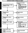
Observations: A man in his late 40s presented with sudden, intense interscapular pain that progressed to paraplegia and sensory loss below T5. He regained neurological function within 6 hours, with residual back pain. Imaging showed SAH and an aneurysm from the left ASA at T5. After a left T4 costotransversectomy, the aneurysm was clipped, and postoperative angiography confirmed ASA patency and aneurysm occlusion. A review of 31 patients (mean [SD] age 43.4 [17.8] years) showed varied treatments: microsurgery (n = 13, 42%), endovascular embolization (n = 3, 10%), conservative management (n = 13, 42%), and surgical exploration followed by conservative management (n = 1, 3%). Complete symptom resolution occurred in 45% (n = 14) of cases.
Lessons: Thoracic ASA aneurysms present diagnostic and treatment challenges. This case illustrates that open microsurgical treatment can successfully decompress the spinal cord and occlude the aneurysm while preserving parent artery flow. https://thejns.org/doi/10.3171/CASE24649.
Keywords: aneurysm; anterior spinal artery; costotransversectomy; microsurgery; spine; subarachnoid hemorrhage; thoracic.
Figures


References
-
- Abdalkader M, Samuelsen BT, Moore JM.Ruptured spinal aneurysms: diagnosis and management paradigms. World Neurosurg. 2021;146:e368-e377. - PubMed
-
- Gonzalez LF Zabramski JM Tabrizi P Wallace RC Massand MG Spetzler RF.. Spontaneous spinal subarachnoid hemorrhage secondary to spinal aneurysms: diagnosis and treatment paradigm. Neurosurgery. 2005;57(6):1127-1131. - PubMed
-
- Madhugiri VS Ambekar S Roopesh Kumar VR Sasidharan GM Nanda A.. Spinal aneurysms: clinicoradiological features and management paradigms. J Neurosurg Spine. 2013;19(1):34-48. - PubMed
-
- Longatti P Sgubin D Di Paola F.. Bleeding spinal artery aneurysms. J Neurosurg Spine. 2008;8(6):574-578. - PubMed
-
- Michon P.. Le Coup de poignard rachidien. Symptôme initial de certaines hémorragies sous-arachnoïdiennes. Essai sur des Hémorragies Méningées Spinales. 1928;36:964-966.
LinkOut - more resources
Full Text Sources

