. 2025 Jun 23;8(1):191.
doi: 10.1038/s42004-025-01568-1.

Developing a new cleavable crosslinker reagent for in-cell crosslinking

Affiliations

Developing a new cleavable crosslinker reagent for in-cell crosslinking

Fränze Müller et al. Commun Chem. .

Abstract

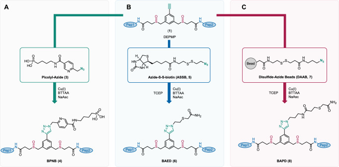
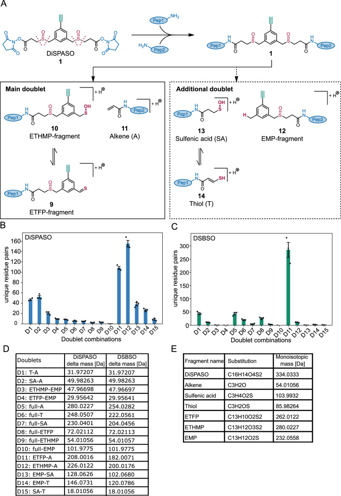
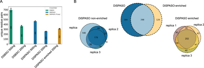
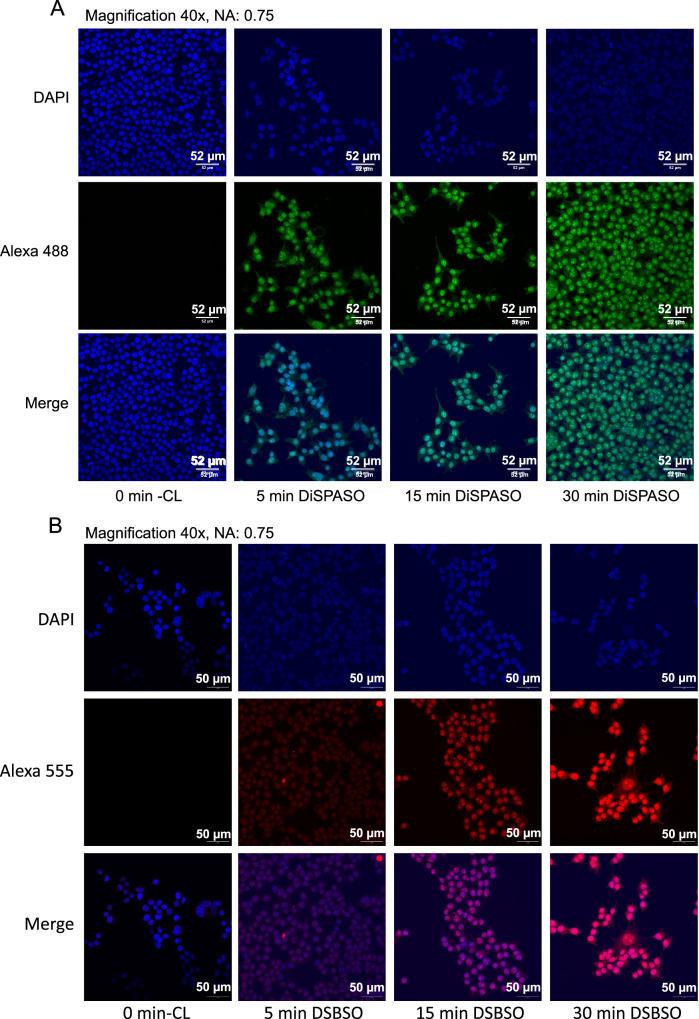
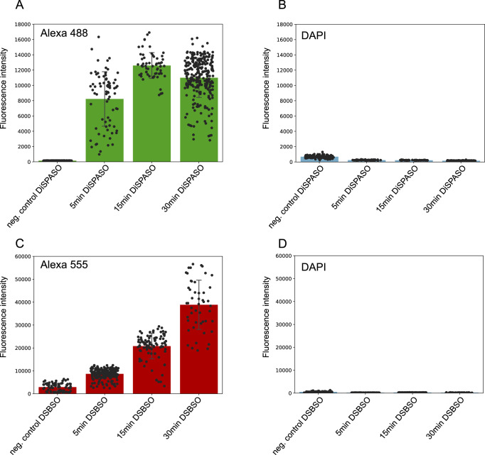
Crosslinking mass spectrometry (XL-MS) is a powerful technology that recently emerged as an essential complementary tool for elucidating protein structures and mapping interactions within a protein network. Crosslinkers which are amenable to post-linking backbone cleavage simplify peptide identification, aid in 3D structure determination and enable system-wide studies of protein-protein interactions (PPIs) in cellular environments. However, state-of-the-art cleavable linkers are fraught with practical limitations, including extensive evaluation of fragmentation energies and fragmentation behavior of the crosslinker backbone. We herein introduce DiSPASO (bis(2,5-dioxopyrrolidin-1-yl) 3,3'-((5-ethynyl-1,3-phenylene)bis(methylenesulfinyl))dipropanoate) as a lysine-selective, MS-cleavable crosslinker with an alkyne handle for affinity enrichment. DiSPASO was designed and developed for efficient cell membrane permeability and crosslinking while securing low cellular perturbation. We tested DiSPASO employing three different copper-based enrichment strategies using model systems with increasing complexity (Cas9-Halo, purified ribosomes, live cells). Fluorescence microscopy in-cell crosslinking experiments revealed a rapid uptake of DiSPASO into HEK 293 cells within 5 minutes. While DiSPASO represents progress in cellular PPI analysis, its limitations and low crosslinking yield in cellular environments require careful optimisation of the crosslinker design, highlighting the complexity of developing effective XL-MS tools and the importance of continuous innovation in accurately mapping PPI networks within dynamic cellular environments.


Conflict of interest statement

Competing interests: The authors declare no competing interests.

Figures

Fig. 1
Fig. 1. Design and synthetic route of the DiSPASO and DiPPASO crosslinkers.
A Design and synthesis of DiSPASO. B Synthetic route: straightforward synthesis of DiPPASO. aSandmeyer Reaction: 1.2 eq. NaNO2/HBr, 0 °C, 1 h, 1.4 eq. CuBr, 0 °C, 18 h. bReduction: 2.0 eq. LiAlH4, THF, 0 °C, 2 h. cBromination: 8.5 eq. PBr3, DCM, 40 °C, 15 h, then H2O, 23 °C, 20 h. dSubstitution: 2.05 eq. methyl 3-mercaptopropanoate, 2.05 eq. K2CO3, DMF, 40 °C, 30 h. eCross-coupling: 1.0 eq. TMS-acetylene, 0.05 eq. Pd(dppf)Cl2-CH2Cl2, 0.05 eq. CuI, THF, 60 °C, 20 h. fGlobal deprotection: 6.0 eq. LiOH, THF/H2O (3:1), 23 °C, 20 h. gNHS/NHP coupling: 2.2 eq. NHS or NHP, 2.2 eq. EDCI-HCl, 0.2 eq. NEt3, DMF, 23 °C, 24 h. hOxidation: 2.0 eq. mCPBA, DCM, 0 °C, 2 h.
Fig. 2
Fig. 2. Topological polar surface area plot of commonly used crosslinker reagents.
DiSPASO is shown in the center of the plot (green dot) with moderate membrane permeability and hydrophobicity compared to other common crosslinker regents. DiPPASO (not presented in this study) shows similar properties to t-BuPhoX (TBDSPP) with a high hydrophobicity compared to other crosslinkers. The clouds of enrichable and permeable crosslinkers are clustered in two distinct parts of the plot, with DiSPASO at the interface between both groups. Classification of the crosslinkers was extracted from the Thermo Scientific crosslinker selection tool. If a crosslinker shows two properties of the classifications the more dominant property was selected for plotting.
Fig. 3
Fig. 3. Overview of DiSPASO enrichment strategies.
A DiSPASO enrichment using a picolyl azide as a click chemistry reagent and an IMAC-based enrichment strategy to retrieve crosslinked peptides from a complex mixture. B Azide-S-S-biotin (ASSB) based enrichment using ASSB as a click chemistry reagent and biotin-streptavidin bead strategy to enrich for crosslinked peptides. The elution of bead-bound crosslinked peptides is performed using the reduction of the disulfide bond of the ASSB compound. C Simplified version of (B). The click reagent Disulfide Azide is already coupled to the beads and the click reaction is taking place directly on the beads. Elution of crosslinked peptides is performed after the click reaction and washing procedure of the beads using a reducing reagent. IUPAC names of all compounds used in this manuscript are described in Supplementary Table S4.
Fig. 4
Fig. 4. Possible fragments of DiSPASO during MS2 fragmentation and evaluation of MS Annika search setting regarding doublet distances.
A Fragmentation products of DiSPASO after high-energy collision dissociation (HCD) showing structures of DSSO-like cleavage products (alkene 11, ETFP 9 and ETHMP-fragments 10, main doublet) and possible additional doublets of the long side of the crosslinker (EMP 12 with SA 13 or T 14 as stubs, dotted gray box). B Numbers of residue pairs identified from Cas9 crosslinked with DiSPASO while searching with each doublet distance separately. D12 (ETHMP-alkene doublet) shows the maximum number of identified links with D11 (ETFP-alkene) as the second and D1, D2 (alkene-thiol, sulfenic acid-alkene) as the third abundant doublets. C Numbers of residue pairs identified from Cas9 crosslinked with DSBSO while searching with each doublet distance separately. D11 (ETFP-alkene doublet) shows the maximum number of identified links. The number of technical replicates is indicated as separate black dots on top of the bar (n = 3). The standard deviation was estimated as average distance from each data point to the sample mean. D Table of doublet definitions and delta masses of all possible doublets. E Table of substitutions and monoisotopic masses of all fragments including DiSPASO as full construct bound to peptides. IUPAC names of all compounds used in this manuscript are described in Supplementary Table S4.
Fig. 5
Fig. 5. Comparison of DiSPASO and DSBSO using Cas9 as a model protein, as well as enrichment performance of DiSPASO using picolyl azide as enrichment tag.
A Number of identified unique crosslinks (unique residue pairs) in a triplicate experiment of Cas9 crosslinked with either DiSPASO or DSBSO in different injection amounts (500 and 200 ng). Blue indicates non-enriched crosslinked peptides, yellow enriched crosslinked peptides from Cas9 DiSPASO experiments. The green color represents Cas9 crosslinking results using DSBSO. The mean value of residue pairs is plotted in the middle of the bar. The number of technical replicates is indicated as separate black dots on top of the bar (n = 3). The standard deviation was estimated as average distance from each data point to the sample mean. B Overlap of identified crosslinks after click reaction and enrichment using picolyl azide as click chemistry reagent. After click reaction and enrichment using picolyl azide the numbers of crosslinks show an overlap of 49% with 21% unique to the enriched crosslinked sample. Left bottom of B: Overlap of technical replicates of non-enriched Cas9 links. Right bottom of B: Overlap of technical replicates of enriched Cas9 links.
Fig. 6
Fig. 6. Application of DiSPASO enrichment strategies in increasing sample complexity.
A Spike-in experiment of crosslinked Cas9 in HeLa background. The Cas9 spike-in amount increased from 0.5 ug to 10 ug in a constant background of 100 ug HeLa. A HeLa in-cell crosslinking sample is used as a “control” sample. The amount of enriched Cas9 links increases with the amount of Cas9 spike-in. B Analysis of Monolinks and linear peptides of the Cas9 spike-in experiment. The Monolinks and linear peptides of the “control” sample show in general fewer peptides due to the different experimental setup of in-cell crosslinking experiments in comparison to a spike-in experiment. The HeLa background peptides of the Cas9 spike-in decrease with increasing enrichable Cas9 crosslinks but cannot be depleted completely. The number of technical replicates is indicated as separate black dots on top of the bar. The standard deviation was estimated as average distance from each data point to the sample mean. C HEK 293 in-cell crosslinking experiment using DiSPASO for crosslinking and Disulfide Azide Agarose beads (DAAB) for click chemistry-based enrichment of crosslinks. The bead amount is increased from 5 uL beads slurry to 120 uL, the control sample is crosslinked with DiSPASO without enrichment. With an increasing number of beads, the number of identifiable crosslinks after enrichment increases. D Analysis of Monolinks and linear peptides of the in-cell crosslinking experiment. Linear peptides could not be depleted after crosslink enrichment.
Fig. 7
Fig. 7. Comparison of DiSPASO and DSBSO uptake during in-cell crosslinking experiments in HEK 293 cells.
A Confocal microscopy images of DiSPASO during in-cell crosslinking experiments with a crosslink duration of 0 min (Control sample without crosslinking, first left panel), 5 min (left second panel), 15 min (third left panel), 30 min (last panel). The nuclei fluorescence signal of DAPI is shown in the upper panel in blue, fluorescence of crosslinked proteins after click reaction to Alexa 488 (green) in the middle panel and a merge of both channels on the bottom. The images were taken on an Olympus Spinning Disk Confocal microscope (2-024) using a magnification of 40 and a numerical aperture of 0.75. B In comparison the DSBSO in-cell experiments were performed in the same way except for the fluorophore. DSBSO has an azide as click reaction handle and therefore Alexa 555 (red) was used to visualize crosslinked proteins. The crosslink duration was set to 0 min (Control sample without crosslinking, first left panel), 5 min (left second panel), 15 min (third left panel), 30 min (last panel). The nuclei fluorescence signal of DAPI is shown in the upper panel in blue, fluorescence of crosslinked proteins after click reaction to Alexa 555 (red) in the middle panel and a merge of both channels on the bottom. The images were also taken on an Olympus Spinning Disk Confocal microscope (2-024) using a magnification of 40 and a numerical aperture of 0.75.
Fig. 8
Fig. 8. Quantitation of crosslink fluorescence signals within nuclei of DiSPASO vs. DSBSO microscopy images.
A Quantitation of the green Alexa 488 signal of DiSPASO crosslinked proteins. The fluorescence intensity increases fast within the first 5 min after reaching a plateau at 30 min. Each dot represents a signal intensity of crosslinked proteins in a nucleus. B Nuclei control signal intensity of DAPI. The background intensity of DAPI is low in comparison to the intensity of crosslinked proteins. C Quantitation of the red Alexa 555 signal of DSBSO crosslinked proteins. The fluorescence intensity increases slowly and reaches its maximum at 30 min. D Nuclei control signal intensity of DAPI. The background intensity of DAPI is also low in comparison to the intensity of crosslinked proteins. Quantitation of signal intensities of all images was performed in Fiji ImageJ (version 1.54f). The number of quantified nuclei is indicated as separate black dots on top of the bar. The standard deviation was estimated as average distance from each data point to the sample mean.
None

References

    1. Tang, X., Wippel, H. H., Chavez, J. D. & Bruce, J. E. Crosslinking mass spectrometry: a link between structural biology and systems biology. Protein Sci.30, 773–784 (2021). - PMC - PubMed
    1. Yu, C. & Huang, L. New advances in cross-linking mass spectrometry toward structural systems biology. Curr. Opin. Chem. Biol.76, 102357 (2023). - PMC - PubMed
    1. Kalathiya, U. et al. Interfaces with structure dynamics of the workhorses from cells revealed through cross-linking mass spectrometry (CLMS). Biomolecules11, 382 (2021). - PMC - PubMed
    1. Piersimoni, L., Kastritis, P. L., Arlt, C. & Sinz, A. Cross-linking mass spectrometry for investigating protein conformations and protein-protein interactions—a method for all seasons. Chem. Rev.122, 7500–7531 (2021). - PubMed
    1. O’Reilly, F. J. & Rappsilber, J. Cross-linking mass spectrometry: methods and applications in structural, molecular and systems biology. Nat. Struct. Mol. Biol.25, 1000–1008 (2018). - PubMed

LinkOut - more resources