. 2025 Jun 23;82(1):252.
doi: 10.1007/s00018-025-05766-x.

PRDM16 acts as a homeostasis regulation factor to suppress the transition of AKI to CKD via upregulation of eukaryotic initiation factor 6

Affiliations

PRDM16 acts as a homeostasis regulation factor to suppress the transition of AKI to CKD via upregulation of eukaryotic initiation factor 6

Xiaozhou Li et al. Cell Mol Life Sci. .

Abstract

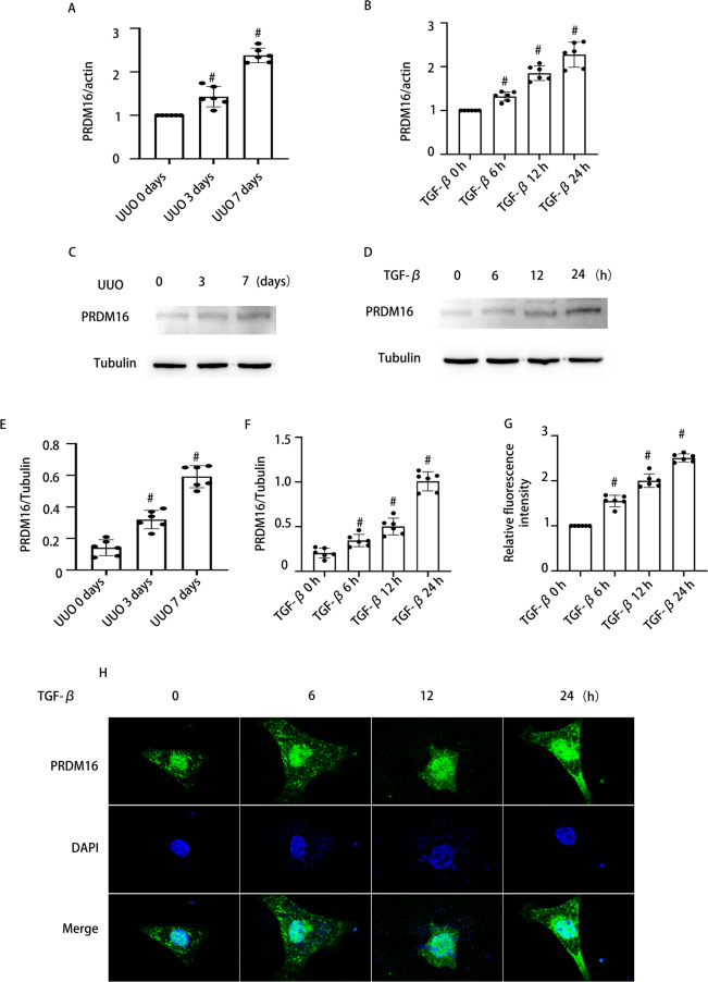
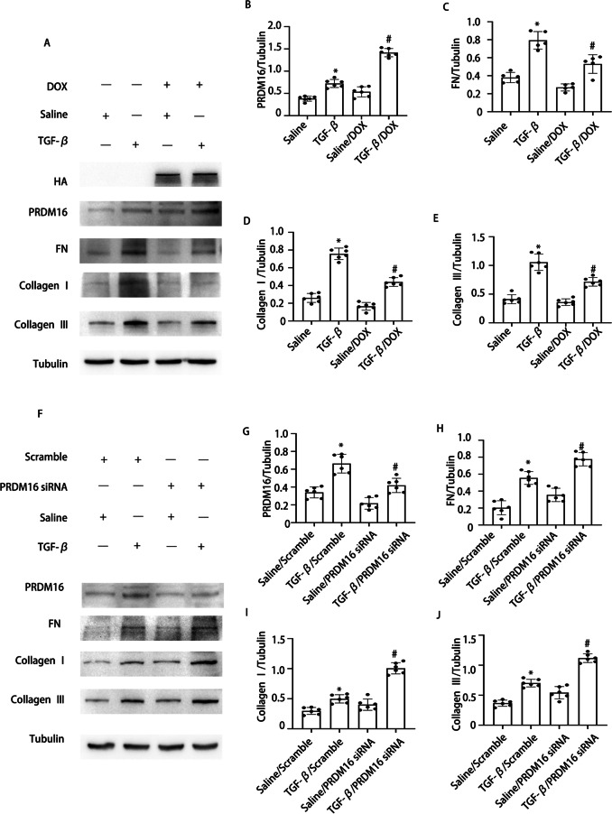
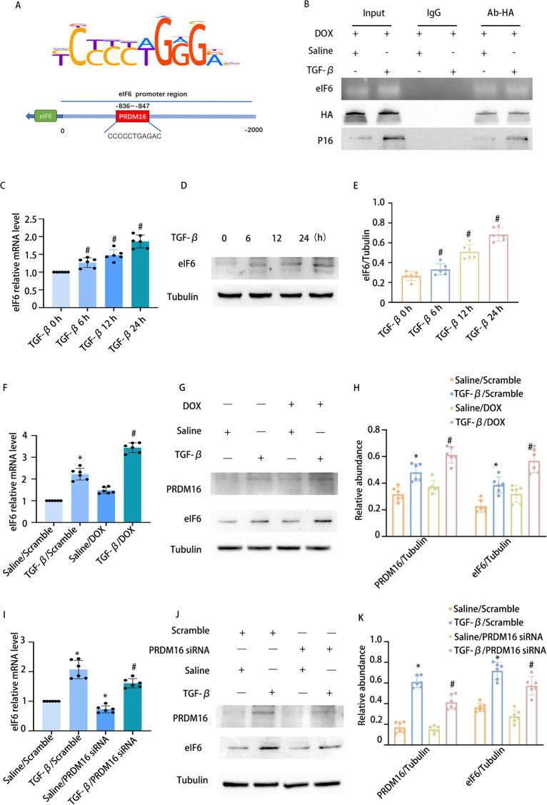
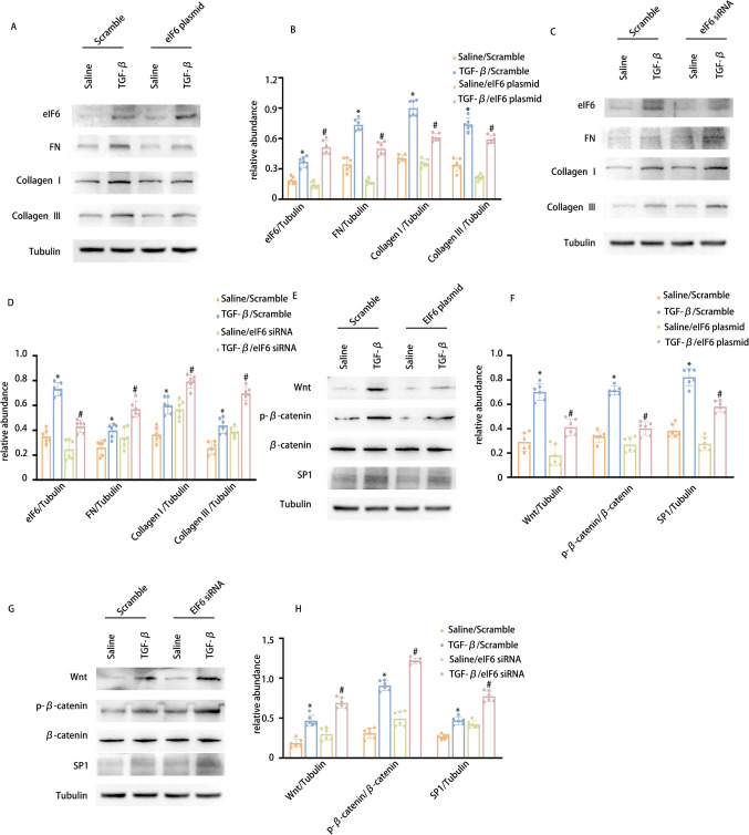
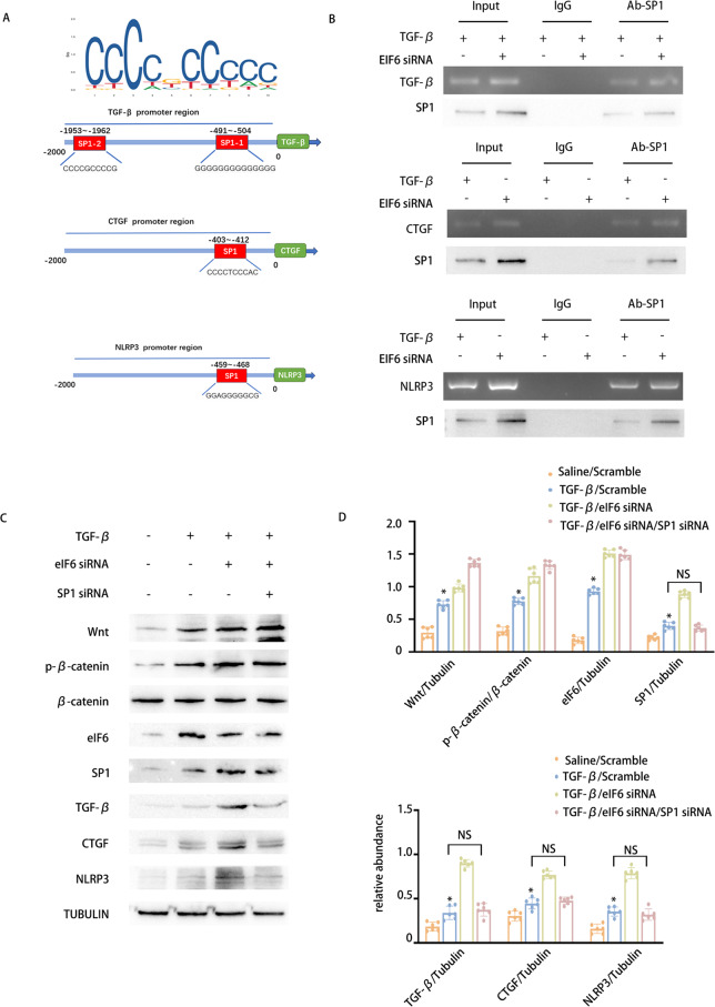
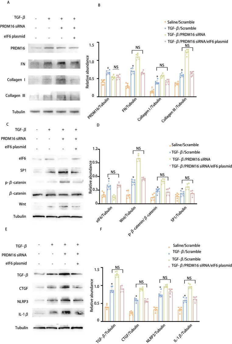
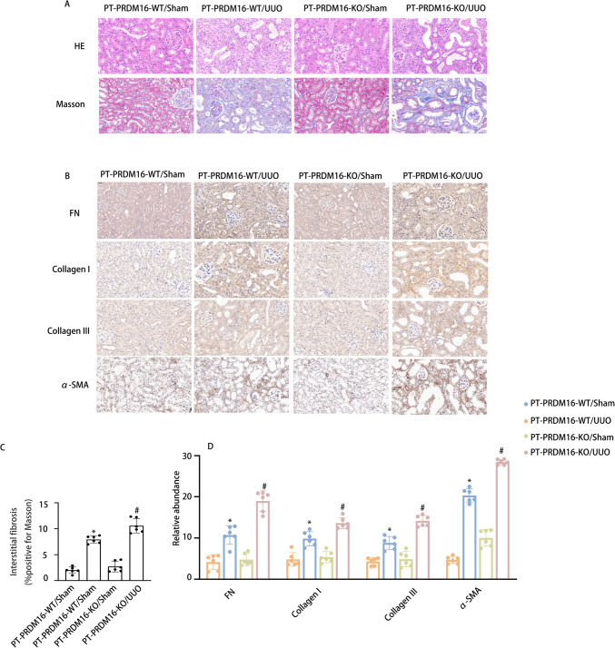
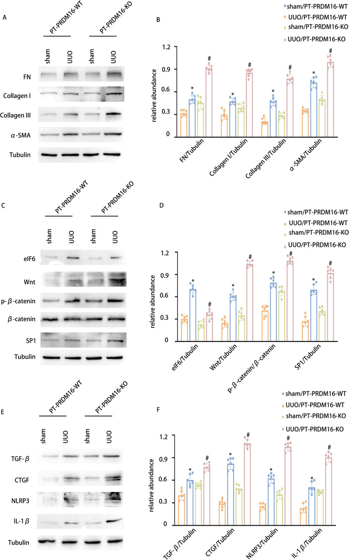
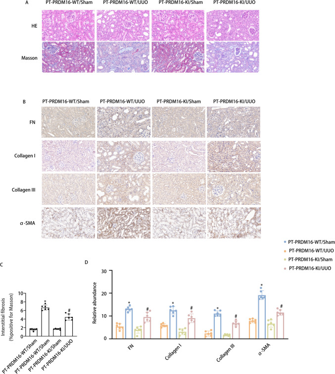
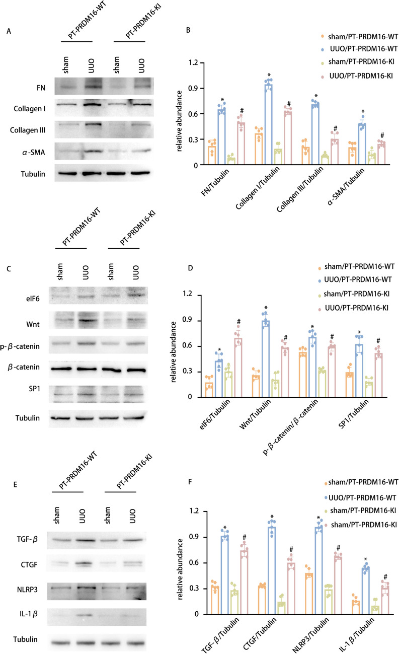
Progressing from acute kidney injury (AKI) to chronic kidney disease (CKD) is acknowledged as a significant clinical challenge. Our recent works indicated that PR domain-containing 16 (PRDM16) impedes the progression of AKI and DKD. Nonetheless, the specific function and regulatory mechanism of PRDM16 during the AKI to CKD transition remain incompletely understood. In this investigation, it was identified that PRDM16 mitigates TGF-β1-induced renal tubulointerstitial fibrosis in BUMPT cells. From a mechanistic perspective, PRDM16 was found to enhance the expression of eif6, which subsequently suppressed TGF-β, CTGF, and NLRP3 levels via the suppression of the Wnt/β-catenin/SP1 signaling cascade. Additionally, knock-in of PRDM16 in kidney proximal tubules resulted in increased expression of eIF6, thereby restraining the stimulation of the Wnt/β-catenin/SP1 pathway, reducing the production of TGF-β, CTGF, and NLRP3, and consequently limiting renal tubulointerstitial fibrosis progression in both unilateral ureteral obstruction and ischemia-reperfusion-injury mouse models.Moreover, overexpression of PRDM16 following ischemia-induced AKI was shown to attenuate renal tubulointerstitial fibrosis and the eIF6/Wnt/β-catenin/SP1/TGF-β, CTGF, and NLRP3 axis. Finally, the PRDM16/eIF6/Wnt/β-catenin/SP1/TGF-β, CTGF, and NLRP3 axis were analyzed in renal biopsies from individuals with minimal change disease and severe obstructive nephropathy. Collectively, these findings indicate that PRDM16-mediated eIF6 induction serves to impede the transition from AKI to CKD by suppressing the Wnt/β-catenin/SP1/TGF-β, CTGF, and NLRP3 axis.

Keywords: AKI; CKD; PRDM16; eIF6.


Conflict of interest statement

Declarations. Conflict of interest: The researchers affirm the absence of any competing interests.

Figures

Fig. 1
Fig. 1
Expression of PRDM16 exposed to TGF-β1 in BUMPT cells and the kidneys of UUO mice and individuals with OB. A-B The mRNA levels of PRDM16, as determined by RT-qPCR, in BUMPT cells with and without TGF-β stimulation, along with the renal tissue of UUO mice. C-D WB of PRDM16 and β-Tubulin in BUMPT cells subjected to TGF-β1 treatment or left untreated, as well as in kidney samples from UUO mice. EF The grayscale analysis (GSY) between them. H Immunofluorescence examination to visualize PRDM16 expression and distribution in BUMPT cells. J Relative fluorescence intensity. Quantification of the PRDM16 -positive cells. Original magnification × 600. Scale bar: 20 µM. Data are denoted as the means ± SD (n = 6). #p < 0.05, vs. control cohort
Fig. 2
Fig. 2
PRDM16 facilitated the TGF-β1-induced expression of fibronectin collagen I and collagen III in BUMPT cells. BUMPT cells underwent transfection with DOX and PRDM16 siRNA, then exposed to 5 ng/ml TGF-β1 or left untreated for 24 h. A WB of PRDM16, FN, HA, collagen I, collagen III, and β-Tubulin in BUMPT cells with or without TGF-β1 and DOX treatment. B-E The GSY between them. F WB of PRDM16, FN, collagen I, collagen III, and β-Tubulin in BUMPT cells with or without TGF-β1 and PRDM16 siRNA treatment. G-J The GSY between them. Representative WB from each cohort (n = 6). Data are denoted as mean ± SD (n = 6). *p < 0.05, TGF-β with Scramble cohort vs. Saline with Scramble cohort; #p < 0.05, TGF-β with DOX cohort or TGF-β with PRDM16 siRNA cohort vs. TGF-β with Scramble cohort
Fig. 3
Fig. 3
PRDM16 facilitated the TGF-β1-induced fibrosis in BUMPT cells via eIF6. BUMPT cells underwent transfection with DOX and PRDM16 siRNA, then exposed to 5 ng/ml TGF-β1 or left untreated for 24 h. A Bioinformatic analysis revealed that the promoter region of the murine eIF6 gene had a putative PRDM16 binding motif. B ChIP assays for PRDM16 were executed utilizing chromatin procured from BUMPT cells. DNA precipitates were amplified using primers flanking the putative PRDM16 binding regions. C eIf6 mRNA levels in BUMPT cells with or without TGF-β treatment were evaluated by RT-qPCR. D eIf6 and β-Tubulin protein expression in BUMPT cells with or without TGF-β1 treatment was examined by WB. E The GSY between them. F eIf6 mRNA expression in BUMPT cells subjected to TGF-β or DOX treatment or left untreated was quantified by RT-qPCR. G WB of eIf6 & PRDM16 and β-Tubulin in BUMPT cells subjected to TGF-β treatment, DOX administration, or left untreated. H The GSY between them. I eIf6 mRNA levels in BUMPT cells subjected to TGF-β treatment, PRDM16 siRNA transfection, or left untreated were quantified by RT-qPCR. J WB of eIf6 & PRDM16 and β-Tubulin in BUMPT cells exposed to TGF-β, PRDM16 siRNA, or left untreated. K The GSY between them. Data are denoted as mean ± SD (n = 6). *p < 0.05, TGF-β with Scramble cohort vs. Saline with Scramble cohort; #p < 0.05, TGF-β with DOX cohort or TGF-β with PRDM16 siRNA cohort vs. TGF-β with Scramble cohort
Fig. 4
Fig. 4
eIF6 modulates SP1 via the Wnt/β-catenin signaling cascade, consequently diminishing the TGF-β1-induced production of collagen I, collagen III, and fibronectin. BUMPT cells underwent transfection with eIf6 plasmid and siRNA, then exposed to 5 ng/ml TGF-β1 or left untreated for 24 h. A WB of eIf6, FN, collagen I, collagen III, and β-Tubulin in BUMPT cells with or without TGF-β1 and eIf6 plasmid treatment. B The GSY between them. C WB of eIf6, FN, collagen I, collagen III, and β-Tubulin in BUMPT cells with or without TGF-β1 and eIf6 siRNA treatment. D The GSY between them. E WB of SP1, Wnt, β-catenin, p-β-catenin, and β-Tubulin in BUMPT cells with or without TGF-β1 and eIf6 plasmid treatment. F The GSY between them. G WB of SP1, Wnt, β-catenin, p-β-catenin, and β-Tubulin in BUMPT cells with or without TGF-β1 and eIf6 siRNA treatment. H The GSY between them Data are denoted as mean ± SD (n = 6). *p < 0.05, TGF-β with Scramble cohort vs. Saline with Scramble cohort; #p < 0.05, TGF-β with eIf6 plasmid cohort or TGF-β with eIf6 siRNA cohort vs. TGF-β with Scramble cohort
Fig. 5
Fig. 5
eIF6 affects the expression of TGF-β、CTGF and NLRP3 via the Wnt/β-catenin/SP1 pathway.The eIf6 and SP1 siRNAs were introduced into BUMPT cells, which were subsequently exposed to 5 ng/ml TGF-β1 or left untreated for 24 h. A Bioinformatic analysis revealed that the promoter region of murine CTGF & TGF-β1 & NLRP3 gene existed a putative SP1 binding motif. B ChIP assays for SP1 were executed with chromatin procured from BUMPT cells. DNA obtained through precipitation was amplified utilizing oligonucleotides encompassing the putative SP1 binding sites for CTGF, TGF-β1, and NLRP3. C WB of eIf6, Wnt, β-catenin, SP1, and β-Tubulin in BUMPT cells subjected to TGF-β1 treatment, eIf6 siRNA, and SP1 siRNA, or left untreated. D The GSY between them. Data are denoted as mean ± SD (n = 6). *p < 0.05, TGF-β1 with Scramble cohort or TGF-β1 with eIf6 siRNA cohort or TGF-β1 with SP1 siRNA cohort vs. Saline with Scramble cohort
Fig. 6
Fig. 6
Inhibition of PRDM16 can reverse the anti-fibrotic effect of eIF6. The eIf6 plasmid and siRNA were introduced into BUMPT cells, which were subsequently exposed to 5 ng/ml TGF-β1 or left untreated for 24 h. A WB of PRDM16, eIf6, FN, collagen I, collagen III, and β-Tubulin in BUMPT cells subjected to TGF-β1 treatment, eIf6 plasmid transfection, and PRDM16 siRNA knockdown. B The GSY between them. Data are denoted as mean ± SD. *p < 0.05, TGF-β with Scramble cohort or TGF-β with PRDM16 siRNA cohort vs. Saline with Scramble cohort. C WB of eIf6, Wnt, β-catenin, SP1, and β-Tubulin in BUMPT cells subjected to TGF-β1 treatment, eIf6 plasmid transfection, and PRDM16 siRNA knockdown. D The GSY between them. E WB of TGF-β1, CTGF, NLRP3, IL-1β, and β-Tubulin in BUMPT cells subjected to TGF-β1 treatment, eIf6 plasmid transfection, and PRDM16 siRNA knockdown. F The GSY between them. Data are denoted as the means ± SD (n = 6). *p < 0.05, TGF-β with eIf6 plasmid cohort or TGF-β with eIf6 siRNA cohort vs. TGF-β with Scramble cohort
Fig. 7
Fig. 7
PRDM16-KO attenuates tubulointerstitial fibrosis in UUO mice. A Hematoxylin–eosin-stained and Representative Masson’s trichrome-stained paraffin-embedded mouse kidney sections. B Semiquantitative scores of tubulointerstitial fibrosis in the kidney cortex. C Immunohistochemical staining of FN, collagen I, collagen III, α-SMA, and F4/80. D Quantification analysis of FN, collagen I, collagen III and α-SMA staining. Original magnification × 400. Scar Bar: 100 µM. Data are denoted as the means ± SD (n = 6). *p < 0.05, UUO with PRDM16-WT cohort vs. sham with PRDM16-WT cohort; #p < 0.05, UUO with PRDM16-KO vs. UUO with PRDM16-WT cohort
Fig. 8
Fig. 8
PRDM16-KO aggravates the expression of FN, Collagen I, collagen III, and α-SMA in UUO mice via/eIF6/Wnt/β-catenin/SP1-axis. A WB of FN, collagen I, collagen III, and α-SMA and β-Tubulin in the cortex of the kidney. B The GSY between them. C WB of eIF6, Wnt, p-β-catenin, β-catenin, SP1, and β-Tubulin in the cortex of the kidney. D The GSY between them. E WB of TGF-β1, CTGF, NLRP3 IL-1β, and β-Tubulin in the cortex of the kidney. F The GSY between them. Data are denoted as the means ± SD (n = 6). *p < 0.05, UUO with PRDM16-WT cohort vs. sham with PRDM16-WT cohort; #p < 0.05, UUO with PRDM16-KO vs. UUO with PRDM16-WT cohort
Fig. 9
Fig. 9
PRDM16-KI alleviated tubulointerstitial fibrosis in UUO mice. A Hematoxylin–eosin-stained and Representative Masson’s trichrome-stained paraffin-embedded mouse kidney sections. B Semiquantitative scores of tubulointerstitial fibrosis in the kidney cortex. C Immunohistochemical staining of FN, collagen I, collagen III, α-SMA, and F4/80. D Quantification analysis of FN, collagen I, collagen III,and α-SMA staining. Original magnification × 400. Scar Bar:100 µM. Data are denoted as the means ± SD (n = 6). *p < 0.05, UUO with PRDM16-WT cohort vs. sham with PRDM16-WT cohort; #p < 0.05, UUO with PRDM16-KI vs. UUO with PRDM16-WT cohort
Fig. 10
Fig. 10
PRDM16-KI attenuates the expression of FN, Collagen I, collagen III, and α-SMA in UUO mice via/eIF6/Wnt/β-catenin/SP1-axis. A WB of FN, collagen I, collagen III, and α-SMA and β-Tubulin in the cortex of the kidney. B The GSY between them. C WB of eIF6, Wnt, p-β-catenin, β-catenin, SP1, and β-Tubulin in the cortex of the kidney. D The GSY between them. E WB of TGF-β1, CTGF, NLRP3 IL-1β, and β-Tubulin in the cortex of the kidney. F The GSY between them. Data are denoted as the means ± SD (n = 6). *p < 0.05, UUO with PRDM16-WT cohort vs. sham with PRDM16-WT cohort; #p < 0.05, UUO with PRDM16-KI vs. UUO with PRDM16-WT cohort

Similar articles

References

    1. Wang S, Qin L (2022) Homeostatic medicine: a strategy for exploring health and disease. Curr Med (Cham, Switzerland) 1(1):16 - PMC - PubMed
    1. Meizlish ML, Franklin RA, Zhou X, Medzhitov R (2021) Tissue homeostasis and inflammation. Annu Rev Immunol 39:557–581 - PubMed
    1. Fries C, Hermiston ML (2023) Challenging T-ALL to IL-7Rp dual inhibition. Blood 142(2):124–126 - PubMed
    1. Puelles VG, Huber TB (2022) Kidneys control inter-organ homeostasis. Nat Rev Nephrol 18(4):207–208 - PMC - PubMed
    1. Tang PM, Nikolic-Paterson DJ, Lan HY (2019) Macrophages: versatile players in renal inflammation and fibrosis. Nat Rev Nephrol 15(3):144–158 - PubMed

MeSH terms

Substances