. 2025 Aug;28(8):1753-1762.
doi: 10.1038/s41593-025-01988-1. Epub 2025 Jun 23.

Closed-loop electrical stimulation prevents focal epilepsy progression and long-term memory impairment

Affiliations

Closed-loop electrical stimulation prevents focal epilepsy progression and long-term memory impairment

Jose J Ferrero et al. Nat Neurosci. 2025 Aug.

Abstract

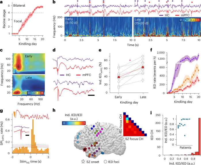
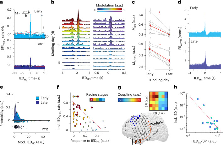
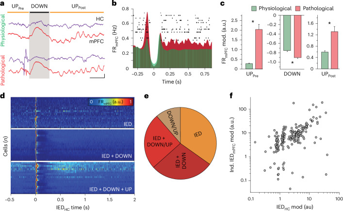
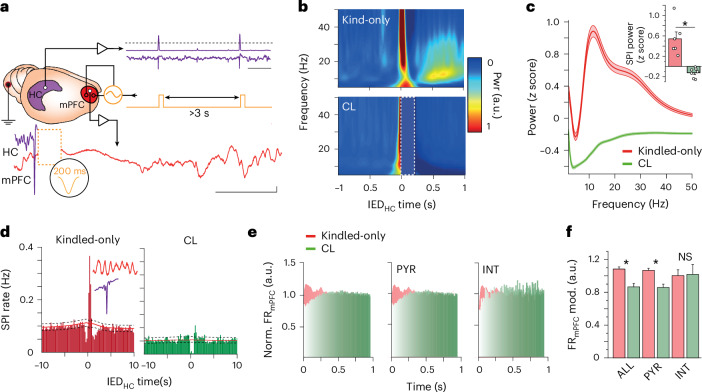
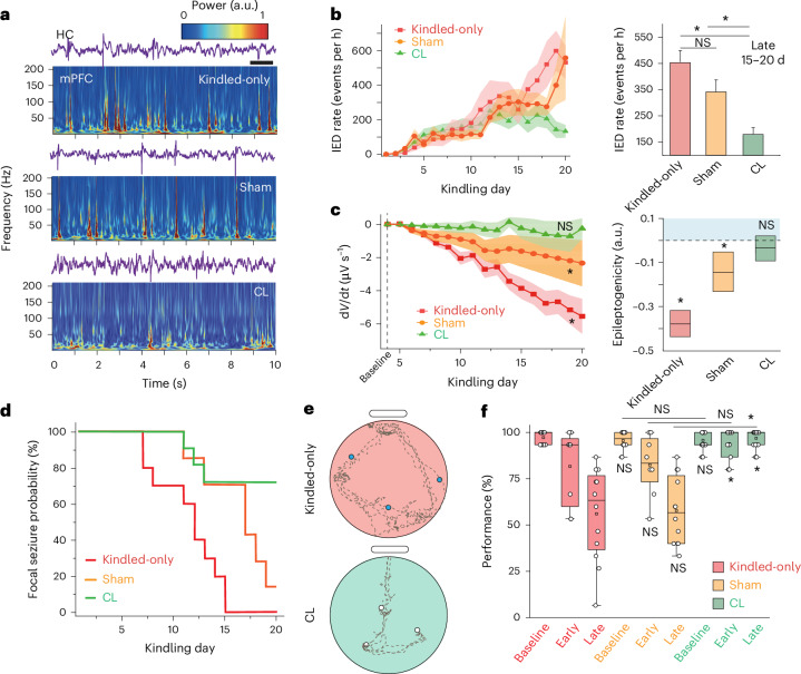
Interictal epileptiform discharges (IEDs) are expressed in epileptic networks and disrupt cognitive functions. It is unclear whether addressing IED-induced dysfunction could improve epilepsy outcomes, as most therapeutic approaches target seizures. We show, in a kindling model of progressive focal epilepsy, that IEDs produce pathological oscillatory coupling associated with prolonged, hypersynchronous neural spiking in synaptically connected cortex and expand the brain territory capable of generating IEDs. A similar relationship between IED-mediated oscillatory coupling and temporal organization of IEDs across brain regions was identified in human participants with refractory focal epilepsy. Spatiotemporally targeted closed-loop electrical stimulation triggered on hippocampal IED occurrence eliminated the abnormal cortical activity patterns, preventing the spread of the epileptic network and ameliorating long-term spatial memory deficits in rodents. These findings suggest that stimulation-based network interventions that normalize interictal dynamics may be an effective treatment of epilepsy and its comorbidities, with a low barrier to clinical translation.


Conflict of interest statement

Competing interests: The authors declare no competing interests.

Figures

Fig. 1
Fig. 1. Progression of focal epilepsy is associated with the creation of an independent focus of interictal epileptic activity.
a, Average Racine stage progression across kindling (n = 10 rats). b, Sample LFP traces acquired simultaneously from HC and mPFC. Dots indicate time of IED (top), with corresponding mPFC spectrogram (bottom) for early and late stages of kindling. c, Sample comodulograms demonstrating decreased cross-frequency coupling between hippocampus and mPFC during progression from early (top) to late stage (bottom) of kindling. d, Sample LFP traces from HC and mPFC, showing IEDHC coupled (top) and independent IEDmPFC (bottom). Scale bar, 100 ms. e, Increase in the percentage of independent mPFC IEDs from early to late stage of kindling (n = 10 rats, 37 early- to 37 late-stage sessions; unpaired two-tailed t-test = −5.34, P = 1.03 × 10−9). *P < 0.05. f, Occurrence of IEDs in hippocampus (purple) and mPFC (total, orange; independent, red) in NREM sleep over kindling (n = 10 rats). g, CCG of a-IEDHC with detected mPFC spindles (bottom) during NREM sleep (95% confidence intervals with midpoints represented as black dashed and red lines, respectively; n = 2,429 spindles, 3,658 stimulations, one sample unkindled rat). Inset, sample mPFC trace after pulse stimulation showing evoked spindle oscillation (top). Independent mPFC (red) IED and HC traces (purple; bottom, scale bar—200 ms). h, Spatial representation of clinically identified IED foci (one color per focus) and seizure onset zone (stars; n = 1 sample human participant). Inset demonstrates the ratio of IEDs in each focus that are independent (>100 ms apart) from seizure onset zone IEDs to total IEDs in the focus (independent IED ratio). Columns represent seizure onset zone channels, and rows represent IED focus channels. i, Histogram of the ratio of independent IEDs to all IEDs across all channels (n = 9 human participants). Inset demonstrates the independent IED ratio across all clinically identified IED foci (n = 9 human participants). ai, Data are presented as mean ± s.e.m. Source data
Fig. 2
Fig. 2. Pathological hippocampal–cortical dynamics link to independent IED foci.
a, Sample CCGs of IEDHC and spindles at early (top) and late (bottom) kindling (early = 1,182/5,737; late = 10,875/4,881 IEDs/SPI). The 95% confidence intervals with midpoints represented as black dashed and red lines, respectively. b, Longitudinal modulation of IEDHC–spindle (right) and hippocampal IEDHC–SO (left) coupling across kindling from a representative rat. c, Decrease in coupling modulation (M) from early to late stage of kindling, for IEDHC–spindle coupling (top; unpaired, two-tailed t-test, 33 early and 29 late sessions, n = 6 rats, t = 5.74, P = 9.3910−7) and IEDHC–SO coupling (bottom; unpaired, two-tailed t-test, 33 early and 29 late sessions, n = 6 rats, t = 7.41, P = 1.31 × 10−9). Data are presented as mean ± s.e.m. *P < 0.05. d, Histogram of clustered mPFC neurons at the time of IEDHC for early (top, n = 69 neurons) and late kindling (n = 110 neurons from sample rat). e, IFR probability distribution of significantly modulated (16.63%) mPFC pyramidal neurons at the time of IEDHC during early and late kindling (unpaired, two-tailed t-test, t  = 5.28, P = 1.00 × 10−7, n = 359 neurons). *P < 0.05. f, Relationship between responsiveness to IEDHC (composite scored based on coupling of IEDHC with SPImPFC, SOmPFC and MUAmPFC) and rate of independent IEDmPFC (n = 57 sessions from six rats; color code indicates Racine stage with dashed line showing pairwise linear correlation; two-tailed Pearson P = 2.80 × 10−5). g, Spatial representation of IED–SPI coupling for two sample IED foci (orange and blue; large circles are IED focus and small circles are maximal region of SPI coupling) from one sample human participant. Inset demonstrates the amount of significant IED–SPI coupling occurring between each IED focus; each column represents an IED focus (n = 6 IED foci from one sample participant). h, Relationship between strength of SPI coupling modulation to seizure onset zone IEDs and rate of IEDs independent from those at seizure onset zone (n = 9 participants). Coupling strengths and IED rates are normalized within participants (two-tailed Spearman r = 0.78; P = 3.30 × 10−7). SO, slow oscillation. Source data
Fig. 3
Fig. 3. Hypersynchronous mPFC neuronal recruitment into pathological oscillatory sequences primes generation of independent mPFC IED-related neural spiking.
a, Sample LFP traces from HC and mPFC demonstrating physiological (top) and pathological (bottom) transition through cortical ‘UP’ and ‘DOWN’ states (scale bar, 500 μV, 200 ms). b, Representative raster plot of mPFC neural spiking (n = 13 sample neurons) overlaid on averaged peri-event firing rate histogram of mPFC neurons during physiological and pathological transition through cortical ‘UP’ and ‘DOWN’ states (n = 351 neurons from one sample rat). c, Differences in mPFC neural firing rate modulation for physiological and pathological transition through cortical ‘UP’ and ‘DOWN’ states—UPpre (left; averaged normalized values for 100 ms preceding cortical ‘DOWN’: Mann–Whitney test, U = 2,289, P = 5.10 × 10−19), DOWN (middle; minimum normalized values for 200 ms following onset of cortical ‘DOWN’ state, U = 11219, P = 4.44 × 10−16), UPpost (right; averaged normalized values for 500 ms after peak of cortical ‘DOWN’ state, U = 5,015, P = 1.85 × 10−4). Data are presented as mean ± s.e.m., and all tests are unpaired, two-tailed with n = 118 sessions. *P < 0.05. d, Representative normalized firing rate heatmap for mPFC neurons modulated by (1) hippocampal IEDs (top), (2) IEDs and pathological coupled cortical ‘DOWN’ state (middle) and (3) IEDs and pathological coupled cortical ‘DOWN’ and ‘UP’ states (bottom); n = 20 most highly modulated neurons per category, derived from five rats. e, Proportion of mPFC neurons modulated by categories of pathological events (n = 617 neurons modulated by pathological events (IED, or pathological IED-induced ‘DOWN/UP’ state) of total 2,733 clustered mPFC neurons from six rats). IED only—34.8%; IED + cortical ‘DOWN’ state—28.4%; IED + cortical ‘DOWN/UP’—25.3%; cortical ‘DOWN’ + cortical ‘UP’ only—11.5%). f, Relationship between mPFC neural firing rate modulation during hippocampal IEDs and independent mPFC IEDs (n = 323 neurons, six rats; two-tailed Spearman ρ = 0.52; P = 3 × 10−24). Source data
Fig. 4
Fig. 4. CL IEDHC-triggered mPFC stimulation prevents pathological cortical response.
a, Schematic representation of CL intervention demonstrating IEDHC detection (scale bar, 1 s), responsively delivered waveform and refractory period between subsequent stimulations. Sample raw mPFC LFP trace after CL stimulation triggered on detected hippocampal IED (stimulation artifact occurs in region of orange dashed box; scale bar = 200 μV, 500 ms). b, Averaged spectrogram at time of IEDHC for a kindled-only (top) and a CL-stimulated (bottom) rat. Dotted box shows the cropped stimulation artifact (n = 1,000 IEDs from sample rat for each condition). c, Sample power spectrum of mPFC LFP following IEDHC (500 ms interval) in kindled-only and CL-stimulated rat (n = 1,000 IEDs from sample rat for each condition). Inset shows change in SPImPFC band power for kindled-only (n = 6 rats) and CL stimulation (from n = 11 rats; unpaired, two-tailed Mann–Whitney, U = 9,580, P = 1.15 × 10−40). *P < 0.05. d, Sample CCG of IEDHC with SPImPFC in a kindled-only rat (left; 8,223 IEDs and 9,974 spindles) and CL-stimulated rat (right; 6,687 IEDs and 5,475 spindles). The 95% confidence intervals with midpoints represented as black dashed and red lines. e, Histogram of all mPFC neuron firing after IEDHC for CL and kindled-only rats (left; CL = 714 neurons, seven rats; kindled-only = 2,733 neurons, six rats), putative pyramidal cells (middle; CL = 587 neurons, seven rats; kindled-only = 2,159 neurons, six rats) and putative interneurons (right; CL = 127 neurons, seven rats; kindled-only = 574 neurons, six rats). f, mPFC neural firing modulation after IEDHC in kindled-only and CL-stimulated rats (quantified during the 500 ms after cortical ‘DOWN’ state for kindled-only rats, and after the end of stimulation for CL-stimulated rats). Kruskal–Wallis with Dunn’s test, χ2 = 36.92—all neurons (P = 2.17 × 10−4, n = 72 and 30 sessions), pyramidal cells (P = 9.71 × 10−4, n = 72 and 30 sessions) and interneurons (P = 1, n = 48 and 27 sessions). *P < 0.05. Data are presented as mean ± s.e.m. in all panels. CL, closed loop; ALL, all neurons; PYR, pyramidal cells; INT, interneurons; NS, not significant. Source data
Fig. 5
Fig. 5. CL stimulation prevents epilepsy progression and memory deterioration.
a, LFP traces from hippocampus and corresponding mPFC spectrograms for sample kindled-only, sham and CL rat. Scale bar, 1 s. b, IEDmPFC occurrence across kindling (left) and quantified at late kindling (right); ANOVA with Bonferroni–Holm correction, P = 2.93 × 10−6, F = 25.47; kindled-only/sham, P = 0.3287; CL/kindled-only; P = 2.64 × 10−6; CL/sham, P = 0.0049; kindled-only n = 39 sessions, 10 rats; sham n = 32 sessions, 7 rats; CL n = 48 sessions, 11 rats. *P < 0.05. c, mPFC epileptogenicity over kindling (left; Mann–Kendall tau; CL, n = 11 rats, P = 0.3; sham, n = 7 rats, P = 8.43 × 10−8; kindled-only, n = 10 rats, P = 8.42 × 10−8). Progression of epileptogenicity over kindling days for kindled-only and sham rats (right; linear mixed-effects model; CL, P = 0.188; sham, P = 0.001; kindled-only, P = 3.51 × 10−26). Box center = coefficient estimate, box boundaries = 5, 95% CI. *P < 0.05. d, Kaplan–Meier curve of progression from focal to bilateral convulsive seizures (kindled-only; n = 10 rats; sham, n = 7 rats; CL, n = 11 rats; log-rank test: kindled-only/sham, P = 1.25 × 10−2; kindled-only/CL, P = 2.42 × 10−3; sham/CL, P = 2.25 × 10−2). e, Examples of exploration path (dashed lines) with reward locations (blue circle, nonretrieved reward; open circle, retrieved reward) for the first three trials of the memory test in late kindling for sample kindled-only and CL rats. f, Memory performance over kindling for kindled-only (n = 4 rats), sham (n = 4 rats) and CL (n = 6 rats): ANOVA with Bonferroni–Holm correction: F = 19.76; kindled-only/sham: baseline (P = 0.77, n = 12/12 sessions), early (P = 0.90, n = 8/8 sessions) and late (P = 0.77, n = 12/12 sessions); sham/CL: baseline (P = 0.98, n = 12/17 sessions), early (P = 0.064, n = 8/11 sessions) and late (P = 4.15 × 10−12, n = 12/17 sessions); kindled-only/CL: baseline (P = 0.77, n = 12/17 sessions), early (P = 0.047, n = 8/11 sessions) and late (P = 7.85 × 10−13, n = 12/17 sessions). *P < 0.05. Data are presented as mean ± s.e.m. unless otherwise noted. Source data

Update of

Similar articles

References

    1. Lagarde, S. et al. Interictal stereotactic-EEG functional connectivity in refractory focal epilepsies. Brain141, 2966–2980 (2018). - PubMed
    1. Hermann, B. P. et al. Cognitive prognosis in chronic temporal lobe epilepsy. Ann. Neurol.60, 80–87 (2006). - PubMed
    1. Kwan, P. & Brodie, M. J. Early identification of refractory epilepsy. N. Engl. J. Med.342, 314–319 (2000). - PubMed
    1. Perrine, K. et al. The relationship of neuropsychological functioning to quality of life in epilepsy. Arch. Neurol.52, 997–1003 (1995). - PubMed
    1. Boylan, L. S. et al. Depression but not seizure frequency predicts quality of life in treatment-resistant epilepsy. Neurology62, 258–261 (2004). - PubMed

LinkOut - more resources