. 2025 Jun 23;22(1):25.
doi: 10.1186/s12950-025-00449-7.

The soluble epoxide hydrolase inhibitor TPPU alleviates Aβ-mediated neuroinflammatory responses in Drosophila melanogaster and cellular models of alzheimer's disease

Affiliations

The soluble epoxide hydrolase inhibitor TPPU alleviates Aβ-mediated neuroinflammatory responses in Drosophila melanogaster and cellular models of alzheimer's disease

Xiaowen Sun et al. J Inflamm (Lond). .

Abstract

Background: Alzheimer's disease (AD) is a common neurodegenerative disease, and its pathogenesis is closely associated with neuroinflammation. The control of neuroinflammation in AD is the focus of current research. soluble epoxide hydrolase (sEH) protein is increased in the brain tissues of patients with AD and has been targeted by multiple genome wide association studies as a prime target for treating AD. Since sEH induces nerve inflammation by degrading epoxyeicosatrienoic acids (EETs), application of sEH inhibitor and sEH gene knockout are effective ways to improve the bioavailability of EETs and inhibit or even resolve neuroinflammation in AD. 1-trifluoromethoxyphenyl-3-(1-propionylpiperidin-4-yl) urea (TPPU) is a potent sEH inhibitor that has been shown to be effective in preclinical animal models of a variety of chronic inflammatory diseases. This study aims to further explore whether TPPU can alleviate AD neuroinflammation.

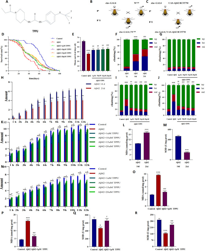
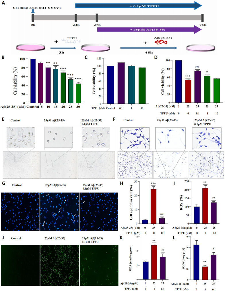
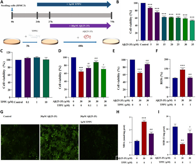
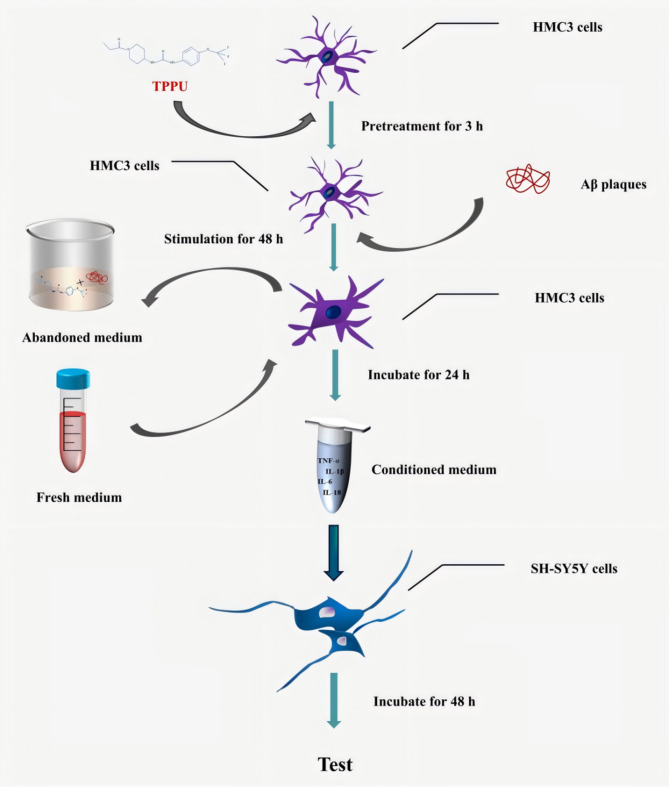
Methods: We established an Aβ42-transgenic Drosophila melanogaster model using the galactose-regulated upstream promoter element 4 (GAL4) / upstream active sequence (UAS) expression system and investigated the protective and anti-neuroinflammatory effects of TPPU against Aβ toxicity. We detected behavioral indexes (survival time, crawling ability, and olfactory memory) and biochemical indexes malondialdehyde (MDA) content and superoxide dismutase (SOD) activity in brain tissues of Aβ42 transgenic flies. Finally, we explored the anti-neuroinflammatory effect of TPPU and its possible mechanism by stimulating cocultures of human SH-SY5Y cells and HMC3 cells with Aβ(25-35) to model neuronal cell inflammation, and evaluated the cells by fluorescence microscopy, ELISA, Western Blot, and Real-time PCR.

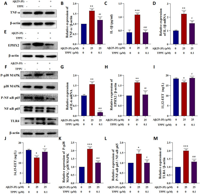
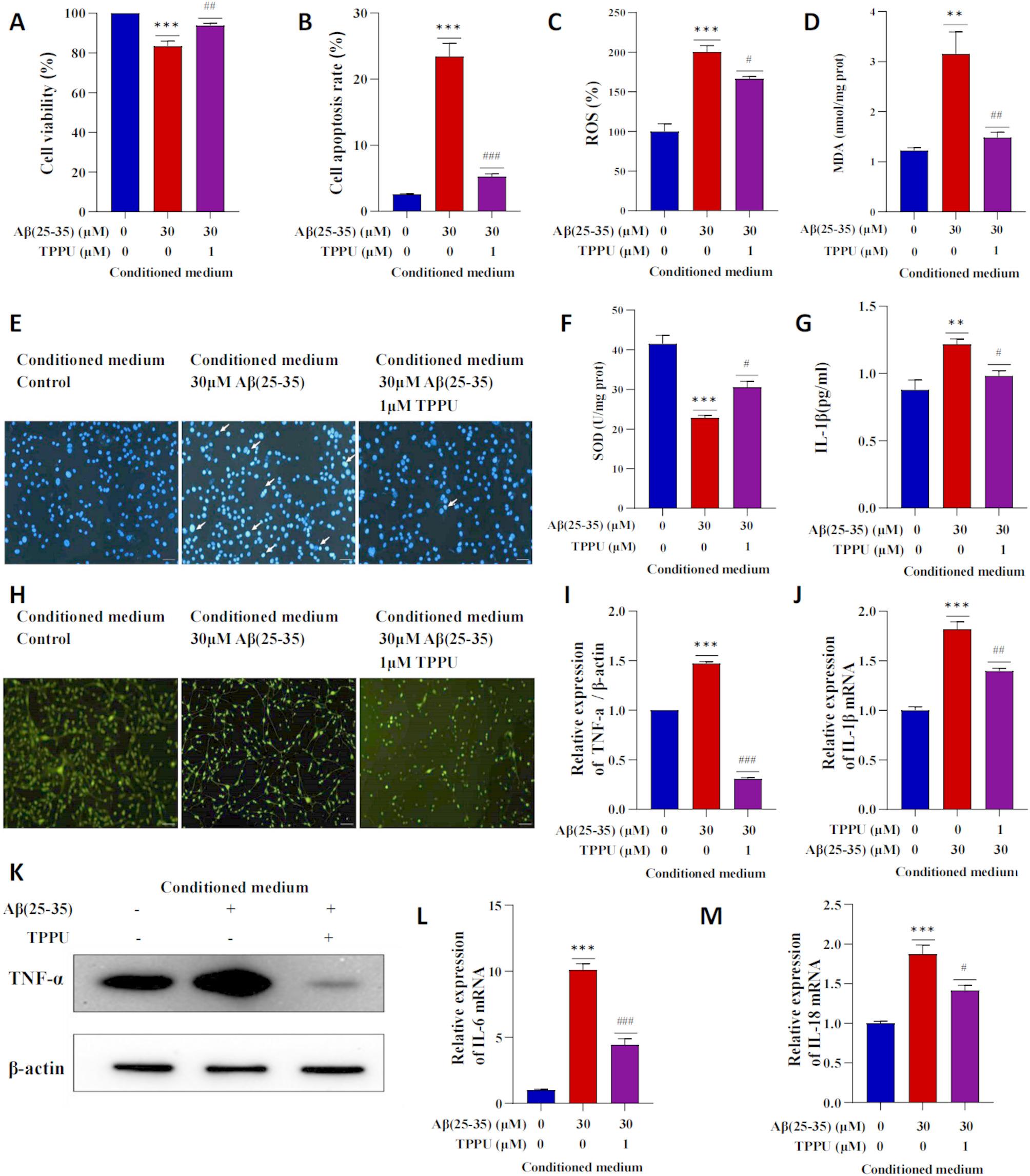
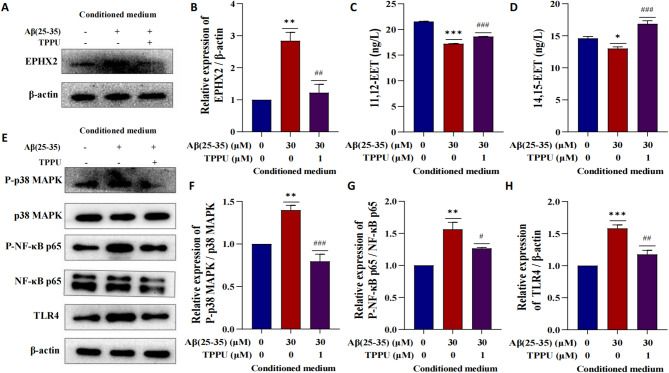
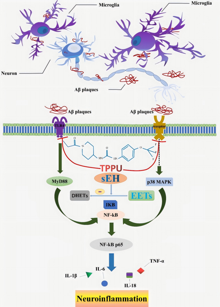
Results: We found that TPPU improved the survival time, crawling ability, and olfactory memory of Aβ42-transgenic flies. We also observed reduction of MDA content and elevation of SOD activity in the brain tissues of these flies. In human cell models, we found that TPPU improved cell viability, reduced cell apoptosis, decreased lipid oxidation, inhibited oxidative damage, thus playing a neuroprotective role. The inflammatory cytokines tumor necrosis factor-α (TNF-α), interleukin-1β (IL-1β), interleukin-6 (IL-6) and interleukin-18 (IL-18) were downregulated, and the mRNA expression of the M2 microglia markers CD206 and SOCS3 were upregulated by TPPU; thus, TPPU inhibited neuroinflammatory responses. TPPU exerted neuroprotective and anti-inflammatory effects by decreasing the protein expression of the sEH-encoding gene EPHX2 and increasing the levels of 11,12-epoxyeicosatrienoic acid (11,12-EET) and 14,15-epoxyeicosatrienoic acid (14,15-EET). The inhibitory effect of TPPU on Aβ(25-35)-mediated neuroinflammation was associated with inhibition of the toll like receptor 4 (TLR4)/nuclear transcription factor-κB (NF-κB) pathway and p38 mitogen activated protein kinases (MAPK)/NF-κB pathway.

Conclusions: We report that the sEH inhibitor TPPU exerts neuroprotective and anti-neuroinflammatory effects in AD models, and it is expected that this drug could potentially be used for the prevention and treatment of AD.

Keywords: Drosophila melanogaster; Alzheimer’s disease; Cell; Neuroinflammation; Neuroprotection; TPPU.


Conflict of interest statement

Declarations. Consent for publication: All listed authors consent to the submission. Competing interests: The authors declare no competing interests.

Figures

Fig. 1
Fig. 1
TPPU improve the behavioral performance and ameliorate oxidative damage of Aβ42-expressing flies. A Chemical structure of TPPU. B Flies cross breed of the control group. C Flies crossbreed of Aβ42-expressing flies’ group. D Flies lifespan curve. E Flies survival time. F Crawling ability of Aβ42-expressing flies at 7 d, 14 d and 21 d of age. ***P<0.001 indicates significant differences between the 7 d and 14 d groups; ###P<0.001 indicates significant differences between the 14 d and 21 d groups. G The crawling ability of 7-d-old flies. H Olfactory memory between Aβ42-expressing flies at 14 d and 21 d of age. *P<0.05, **P<0.01 and ***P<0.001. I The crawling ability of 14-d-old flies. J The crawling ability of 21-d-old flies. K Olfactory memory experiment with flies at 14 d of age. L MDA content in brain tissue of Aβ42-expressing flies at 14 d and 21 d of age. ***P<0.001. M SOD activity in brain tissue of Aβ42-expressing flies at 14 d and 21 d of age. ***P<0.001. N Olfactory memory experiment of flies at 21 d of age. O MDA content in brain tissues of 14-d-old flies. P MDA content in brain tissues of 21-d-old flies. Q SOD activity in brain tissues of 14-d-old flies. R SOD activity in brain tissues of 21-d-old flies. E, G, I-K, N-R *P<0.05, **P<0.01 and ***P<0.001 indicate significant differences in Aβ42-expressing flies group compared with the control group. #P<0.05, ##P<0.01 and ###P<0.001 indicate significant differences in Aβ42-expressing flies group compared with the TPPU-treated group. n=3, means ± SEMs. The data were analyzed using Student's t test or one-way analysis of variance (ANOVA) and Bonferroni post event test
Fig. 2
Fig. 2
TPPU reduces Aβ(25–35)-mediated neurotoxicity and oxidative damage in SH-SY5Y cells. A SH-SY5Y cells experimental design. B The effect of different concentrations of Aβ(25–35) on the viability of SH-SY5Y cells was determined for 48 h by CCK-8. C Assessment of TPPU-induced cytotoxicity in SH-SY5Y cells. SH-SY5Y cells were treated with TPPU at different concentrations (0.1 µM, 1 µM or 10 µM) for 48 h. D The effect of TPPU pretreatment on the viability of SH-SY5Y cells after treatment with Aβ(25–35). Cells were pretreated with varying concentrations of TPPU for 3 h followed by 25 µM Aβ(25–35) stimulation for 48 h. E Morphological changes in SH-SY5Y cells under the microscope. Scale bar = 100 μm. F The morphological changes of SH-SY5Y cells after crystal violet staining. Scale bar = 100 μm. G Hoechst 33258 staining; arrow indicates apoptotic cells. Scale bar = 100 μm. H Quantitative analysis of apoptotic cells that were identified by Hoechst 33258 staining. I Verage fluorescence intensity of ROS (normalized to the control group). J ROS were imaged under a fluorescence microscope; green fluorescence represents the fluorescence intensity of the ROS probe DCFH-DA. Scale bar = 100 μm. K MDA content. L SOD activity. The Aβ(25–35) group was compared with the control group, *P < 0.05, **P < 0.01 and ***P < 0.001; the Aβ(25–35) group was compared with the TPPU pretreatment group, #P < 0.05, ##P < 0.01 and ###P < 0.001. n = 3, means ± SEMs. The data were analyzed using one-way analysis of variance (ANOVA) and Bonferroni post event test
Fig. 3
Fig. 3
TPPU against Aβ(25–35)-mediated neuroinflammatory in SH-SY5Y cells. The expression levels of TNF-α (A, B), EPHX2 (E, H), P-p38 MAPK (F, K), p38 MAPK (F), P-NF-κB p65 (F, L), NF-κB p65 (F) and TLR4 (F, M) in SH-SY5Y cells were analyzed by Western Blot. IL-1β (C), 11,12-EET (I) and 14,15-EET (J) levels in cell culture medium were measured by ELISA. The mRNA expression of IL-1β (D) and IL-6 (G) were measured by real-time PCR. The Aβ(25–35) group was compared with the control group, *P < 0.05, **P < 0.01 and ***P < 0.001; the Aβ(25–35) group was compared with the TPPU group, #P < 0.05, ##P < 0.01 and ###P < 0.001. n = 3, means ± SEMs. The data were analyzed using one-way analysis of variance (ANOVA) and Bonferroni post event test
Fig. 4
Fig. 4
TPPU reduces Aβ(25–35)-induced oxidative damage in HMC3 cells. A Schematic of the HMC3 cell experimental design. B The effect of different concentrations of Aβ(25–35) on the viability of HMC3 cells after 48 h was determined by CCK-8 assay. C Assessment of TPPU cytotoxicity in HMC3 cells treated with different concentrations of TPPU (0.1 µM, 1 µM or 10 µM) for 48 h. D The effect of TPPU pretreatment on the viability of HMC3 cells after treatment with Aβ(25–35). HMC3 cells were pretreated with varying concentrations of TPPU for 3 h followed by 30 µM Aβ(25–35) stimulation for 48 h. E HMC3 cells were pretreated with 1 µM TPPU for 3 h and then stimulated with 30 µM Aβ(25–35) for 48 h. F Average fluorescence intensity of ROS (normalized to the control group). G ROS were imaged under a fluorescence microscope. Green fluorescence represents ROS that were labeled with the ROS probe DCFH-DA. Scale bar = 100 μm. H MDA content. I SOD activity. The Aβ(25–35) group was compared with the control group, *P < 0.05, **P < 0.01 and ***P < 0.001; the Aβ(25–35) group was compared with the TPPU pretreatment group, #P < 0.05, ##P < 0.01 and ###P < 0.001. n = 3, means ± SEMs. The data were analyzed using one-way analysis of variance (ANOVA) and Bonferroni post event test
Fig. 5
Fig. 5
TPPU against the Aβ(25–35)-induced neuroinflammatory response in HMC3 cells. The expression levels of TNF-α (A, B), EPHX2 (I, K), P-p38 MAPK (M, O), p38 MAPK (M), P-NF-κB p65 (M, P), NF-κB p65 (M), MyD88 (M, Q) and TLR4 (M, R) in HMC3 cells were analyzed by Western Blot. IL-1β (C), 11,12-EET (L) and 14,15-EET (N) levels in cell culture medium were measured by ELISA. The mRNA expression of TNF (D), IL-1β (E), IL-6 (F), IL-18 (G), SOCS3 (H) and CD206 (J) were measured by real-time PCR. The Aβ(25–35) group was compared with the control group, *P < 0.05, **P < 0.01 and ***P < 0.001; the Aβ(25–35) group was compared with the TPPU pretreatment group, #P < 0.05, ##P < 0.01 and ###P < 0.001. n = 3, means ± SEMs. The data were analyzed using one-way analysis of variance (ANOVA) and Bonferroni post event test
Fig. 6
Fig. 6
Schematic diagram of coculturing experiments. HMC3 cells were pretreated with TPPU for 3 h followed by Aβ(25–35) stimulation for an additional 48 h. The medium was replaced with fresh medium and then incubated for 24 h to prepare conditioned medium containing various inflammatory cytokines secreted by HMC3 cells. The collected conditioned medium was centrifuged, added to SH-SY5Y cells, and incubated for 48 h for further analysis
Fig. 7
Fig. 7
The neuroprotective and anti-neuroinflammatory effects of TPPU on SH-SY5Y cells cultured with conditioned medium from HMC3 cells. A Viability was determined by CCK-8 assay. B Apoptotic cells were quantified by Hoechst 33258 staining. C Average fluorescence intensity of ROS (normalized to the control group). D MDA content. E Hoechst 33258 staining; arrows indicate apoptotic cells. Scale bar=100 μm. F SOD activity. G IL-1β levels in cell culture medium were measured by ELISA. H ROS were imaged under a fluorescence microscope. I, K The expression levels of TNF-α in SH-SY5Y cells cultured with conditioned medium from HMC3 cells was analyzed by Western Blot. The mRNA expression of IL-1β (J), ILL-6 (L), IL-18 (M) were measured by real-time PCR. SH-SY5Y cells cultured with medium from the Aβ group were compared with SH-SY5Y cells cultured with control medium, *P<0.05, **P<0.01 and ***P<0.001; SH-SY5Y cells cultured with medium from the Aβ group were compared with SH-SY5Y cells cultured with medium from the TPPU group, #P<0.05, ##P<0.01 and ###P<0.001. n=3, means ± SEMs. The data were analyzed using one-way analysis of variance (ANOVA) and Bonferroni post event test
Fig. 8
Fig. 8
TPPU exerts an antineuroinflammatory effect by regulating the TLR4/NF-κB and p38 MAPK/NF-κB signaling pathways in SH-SY5Y cells cultured with conditioned medium from HMC3 cells. The expression levels of EPHX2 (A, B), P-p38 MAPK (E, F), p38 MAPK (E), P-NF-κB p65 (E, G), NF-κB p65 (E), and TLR4 (E, H) in SH-SY5Y cells cultured with conditioned medium from HMC3 cells were analyzed by Western Blot. 11,12-EET (C) and 14,15-EET (D) levels in cell culture medium were measured by ELISA. SH-SY5Y cells cultured with medium from the Aβ group were compared with SH-SY5Y cells cultured with control medium, *P < 0.05, **P < 0.01 and ***P < 0.001; SH-SY5Y cells cultured with medium from the Aβ group were compared with SH-SY5Y cells cultured with medium from the TPPU group, #P < 0.05, ##P < 0.01 and ###P < 0.001. n = 3, means ± SEMs. The data were analyzed using one-way analysis of variance (ANOVA) and Bonferroni post event test
Fig. 9
Fig. 9
Potential mechanism underlying the protective effect of TPPU against neuroinflammation in Alzheimer’s disease

Similar articles

References

    1. Serrano-Pozo A, Frosch MP, Masliah E, Hyman BT. Neuropathological alterations in alzheimer disease. Cold Spring Harbor Perspect Med. 2011;11:a006189. - PMC - PubMed
    1. Scheltens P, De Strooper B, Kivipelto M, Holstege H, Chételat G, Teunissen CE, et al. Alzheimer’s disease. Lancet (London England). 2021;39710284:1577–90. - PMC - PubMed
    1. Estimation of the global prevalence of dementia. In 2019 and forecasted prevalence in 2050: an analysis for the global burden of disease study 2019. Lancet Public Health. 2022;72:e105–25. - PMC - PubMed
    1. Ferrari C, Sorbi S. The complexity of alzheimer’s disease: an evolving puzzle. Physiol Rev. 2021;1013:1047–81. - PubMed
    1. Tatulian SA. Challenges and hopes for alzheimer’s disease. Drug Discovery Today. 2022;274:1027–43. - PubMed

LinkOut - more resources