. 2025 Jun 24;31(1):247.
doi: 10.1186/s10020-025-01264-9.

Epigenetic modulation elicits an NK cell-mediated immune response in urothelial carcinoma

Affiliations

Epigenetic modulation elicits an NK cell-mediated immune response in urothelial carcinoma

Himani Kumari et al. Mol Med. .

Abstract

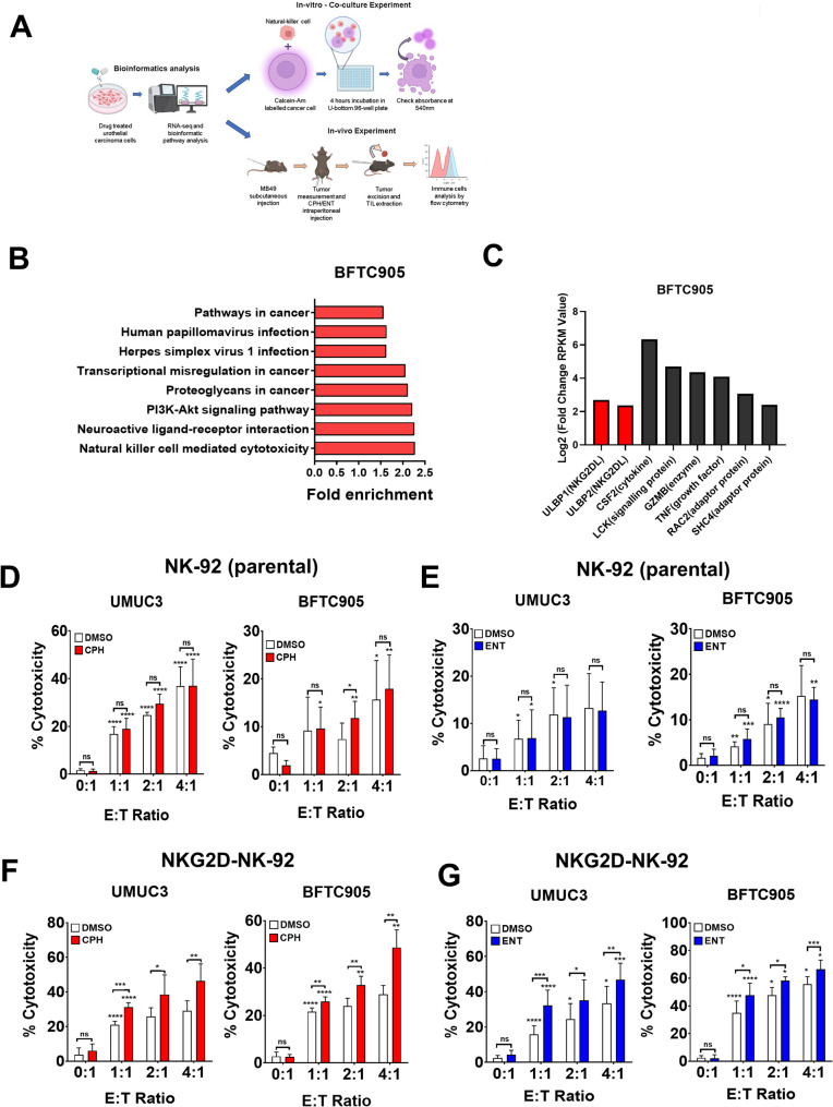
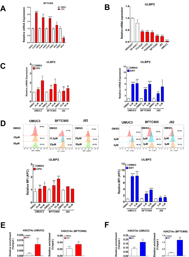
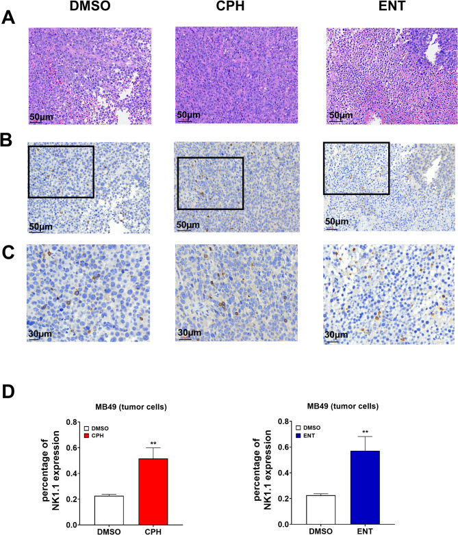
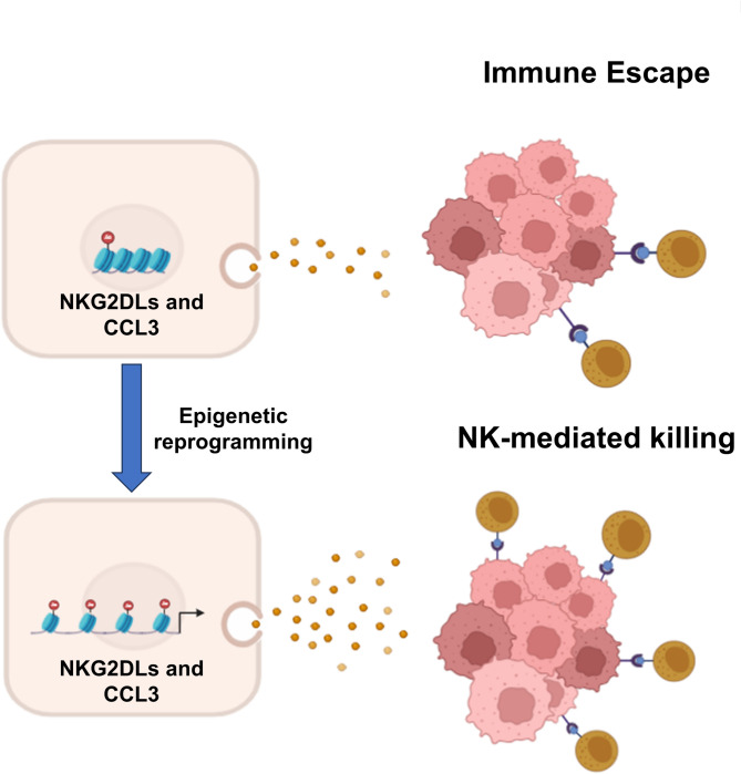
Urothelial carcinoma (UC), the second most prevalent cancer of the urothelial system, has demonstrated a well-developed ability to evade natural killer (NK) cell-mediated killing via various pathways, highlighting the need for innovative therapeutic approaches. Our study examines the role of cyproheptadine (CPH) and entinostat (ENT) as epigenetic modifiers in enhancing NK cell-mediated cytotoxicity against UC. Through our in vitro experiments, we observed that pre-treatment with CPH or ENT significantly increased NK cell-mediated elimination of UC cells, as demonstrated in NK cytotoxicity assay wherein UC cells were co-cultured with both NK-92 and human primary NK cells. This enhancement is attributed to the epigenetic activation of NKG2D ligands, notably through increased H3K27ac enrichment and reduced enrichment of H3K27me3 at the ULBP2 promoter region in UC cells. Additionally, ectopic expression of ULBP2 in UC cells further augmented their susceptibility to NK cell-mediated killing. Importantly, our syngeneic mouse model using MB49 tumor cells, showed that CPH or ENT treatments significantly reduced tumor growth via promoting NK cell infiltration into the tumor microenvironment. This increased infiltration is likely due to elevated levels of the NK-recruiting chemokine CCL3 in drug-treated UC cells. Collectively, these results underscore the potential of CPH and ENT to suppress tumor growth and enhance immune surveillance by modulating epigenetic pathways that boost NK cell activity, offering promising insights into new strategies for UC treatment.

Supplementary Information: The online version contains supplementary material available at 10.1186/s10020-025-01264-9.

Keywords: CCL3; CPH; Innate immunity; NK cells; NKG2DL; ULBP2; Urothelial carcinoma.


Conflict of interest statement

Declarations. Ethics approval and consent to participate: This study was approved by the Institutional Review Board (IRB) of Taoyuan General Hospital (IRB no.: TYGH109026), Ministry of Health and Welfare and Institutional Animal Care and Use committee (IACUC) of the National Chung Cheng University, Taiwan. Consent for publication: All authors read the final manuscript and agreed to publish it. Competing interests: The authors declare no competing interests.

Figures

Fig. 1
Fig. 1
NKG2D NK-92 cells exhibited enhanced NK cell-mediated cytotoxicity in UC cells pretreated with epigenetic drug. A schematic representation of the experimental setup. B Histogram showing enrichment of upregulated genes associated pathways and C list of upregulated genes using RNA-Seq data from BFTC905 UC cells treated with cyproheptadine (CPH). UMUC3 and BFTC905 cells pretreated cyproheptadine (CPH, 50 µM in UMUC3 and 25 µM in BFTC905 cells) or Entinostat (ENT, 2 µM) were co-cultured with either (D, E) parental NK-92 or (F, G) NKG2D-NK-92 cells. Bar chart showing cell specific lysis with different effector to target (E: T) ratio as the measure of NK cell-mediated cytotoxicity via calcein-AM assay (For detail, please see material and methods). Interestingly, pretreatment with CPH or ENT only further enhanced the NK cell-mediated cytotoxicity in NKG2D-NK-92 cells (F, G), but not parental NK-92 cells (D, E). Each error bar represents mean ± SD from triplicates. The significance is calculated by unpaired T-test. ****P < 0.0001; *** P < 0.0005; **P < 0.01; *P < 0.05. Individual asterisk shown on each bar compares to the previous E to T ratio bar of each group
Fig. 2
Fig. 2
Primary human NK cells exhibited enhanced NK cell-mediated cytotoxicity in UC pretreated with epigenetic drug. Primary NK cells were isolated from cord blood of three health donor. A Representative flow cytometry analysis showing purity of expanded primary human NK cells using CD3−veCD56+ve. Left panel: lymphocyte population; middle panel: CD3−ve population; right panel: CD3−veCD56+ve population. Bar chart showing cell specific lysis with different E:T ratio as the measure of NK cell-mediated cytotoxicity using expanded primary NK cells, via calcein-AM assay, in BFTC905 cells pretreated with (B) 25 µM CPH or (C) 2 µM ENT. Representative experiments of human primary NK cells isolated from the cord blood PBMCs of one healthy donor’s were shown. Each error bar represents mean ± SD from triplicates. The significance is calculated by unpaired T-test. ****P < 0.0001; *** P < 0.0005; **P < 0.01; *P < 0.05. Individual asterisk shown on each bar compares to the previous E to T ratio bar of each group
Fig. 3
Fig. 3
Restoration of NKG2D ligands in urothelial carcinoma treated with CPH or ENT. A Bar chart showing the relative mRNA expression of NKG2DLs in BFTC905 cells treated with CPH 25µM for 48 h using qRT-PCR. B Relative expression of ULBP2 in HEK293T, SV-HUC1 and a panel of UC cell lines by quantitative RT-PCR. Expression of ULBP2 in UC cells treated with CPH (left) or ENT (right) by (C) quantitative RT-PCR and (D) flow cytometry analysis. Enrichment of H3K27 Ac at the promoter of ULBP2 was determined in UC cells treated with (E) CPH (25 µM in BFTC905, 50µM in UMUC3) or (F) ENT (2µM) as determined by ChIP-qPCR. Each error bar represents mean ± SD from triplicates. The significance is calculated by unpaired T-test. ****P < 0.0001; *** P < 0.0005; **P < 0.01; *P < 0.05
Fig. 4
Fig. 4
Enhanced NK cell-mediated cytotoxicity in ULBP2 overexpressing UC cells. Flow cytometry analysis of UMUC3 cells expressing (A) control, (B) low, or (C) high level of ULBP2, as determined by ULBP2 (middle panel) and GFP intensity (right panel). D Bar chart showing cell specific lysis via calcein-AM assay with different E:T ratio of NKG2D-NK-92 cells vs. UMUC3 with different expression level of ULBP2 (white: control, no expressing; blue, low expression; red: high expression) in BFTC905 cells. Each error bar represents mean ± SD from triplicates. The significance is calculated by unpaired T-test. ****P < 0.0001; *** P < 0.0005; **P < 0.01; *P < 0.05. Individual asterisk shown on each bar compares to the previous E to T ratio bar of each group
Fig. 5
Fig. 5
Epigenetic treatment delays tumor progression in syngeneic tumor mice model. A Schematic representation of the treatment plan. In brief, MB49 UC cells were injected subcutaneously into C57BL/6 mice. Eight days after the tumor inoculation, 5 mg/kg of CPH or 3 mg/kg of ENT were injected i.p. 5 days per week for 2 weeks. Tumor volume was measured daily. Tumor volume of mice treated with (B) CPH (n = 7/group) or (C) ENT (n = 4/group) was shown. Dot plot showing tumor weight, infiltrated CD3-ve NK and CD3 + ve NKT cells, as determined by flow cytometry, in mice treated with (D) CPH or (E) ENT, respectively. The significance is calculated by unpaired T-test. *P < 0.05
Fig. 6
Fig. 6
Infiltration of NK cells in tumor microenvironment of syngeneic tumor mice model. A Representational illustration of HE stained sample of tumor tissues. B IHC stained of NK1.1 showing infiltrated NK cells in tumor microenvironment in dark brown colored cells. The boxed region is enlarged and shown in (C). D Bar graph showing percentage of infiltrated NK cells in tumor microenvironment. Infiltrated cells were determined using imageJ software. The significance was calculated by unpaired T-test. **P < 0.01
Fig. 7
Fig. 7
CPH treatment increases the expression of CCL3 in UC cells. A Histogram showing expression of chemokines in MB49 cells treated with CPH as determined by RNA-Seq. B Volcano plot representing differentially expressed genes of MB49 cells treated with CPH. C Venn diagram representing differential upregulated genes and 12 common genes in MB49 cells and BFTC905 cells treated with CPH. Relative expression level of CCL3 mRNA in (D, E) human and (F, G) mice UC cells treated with (D, F) CPH or (E, G) ENT, as determined by quantitative RT-PCR. Relative protein expression of CCL3 in MB49 parental cells treated with (H) CPH and (I) ENT is quantified by ELISA. Each error bar represents mean ± SD from triplicates. The significance is calculated by unpaired T-test. ****P < 0.0001; *** P < 0.0005; **P < 0.01; *P < 0.05
Fig. 8
Fig. 8
The schematic model depicts that CPH or ENT treatment reprograms the epigenome resulting in the upregulation of NKG2D ligands and CCL3 to enhance the NK cell-mediated cytotoxicity in UC

References

    1. Adamik J, et al. EZH2 or HDAC1 Inhibition reverses multiple Myeloma-Induced epigenetic suppression of osteoblast differentiation. Mol Cancer Res. 2017;15:405–17. - PMC - PubMed
    1. Allen F, et al. CCL3 augments tumor rejection and enhances CD8(+) T cell infiltration through NK and CD103(+) dendritic cell recruitment via IFNgamma. Oncoimmunology. 2018;7:e1393598. - PMC - PubMed
    1. Arai S, et al. Infusion of the allogeneic cell line NK-92 in patients with advanced renal cell cancer or melanoma: a phase I trial. Cytotherapy. 2008;10:625–32. - PubMed
    1. Armeanu S, et al. Natural killer cell-mediated Lysis of hepatoma cells via specific induction of NKG2D ligands by the histone deacetylase inhibitor sodium valproate. Cancer Res. 2005;65:6321–9. - PubMed
    1. Bauman Y, et al. Downregulation of the stress-induced ligand ULBP1 following SV40 infection confers viral evasion from NK cell cytotoxicity. Oncotarget. 2016;7:15369–81. - PMC - PubMed

LinkOut - more resources