. 2025 Jun 23;26(1):121.
doi: 10.1186/s40360-025-00947-7.

Naringin mitigated doxorubicin-induced kidney injury by the reduction of oxidative stress and inflammation with a synergistic anticancer effect

Affiliations

Naringin mitigated doxorubicin-induced kidney injury by the reduction of oxidative stress and inflammation with a synergistic anticancer effect

Nahla S Gad et al. BMC Pharmacol Toxicol. .

Abstract

Background: The pathophysiology and severity of kidney impairment due to doxorubicin (DOX) treatment are markedly influenced by oxidative stress and inflammation. Naringin (NG), a natural flavonoid, has anti-inflammatory and antioxidant properties. The nephroprotective effect of NG on DOX-induced kidney toxicity was investigated to increase its utility in clinical settings.

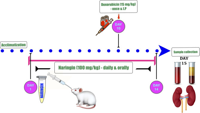
Methods: DOX toxicity was induced by a single ip injection (15 mg/kg) and for possible protection NG (100 mg/Kg) was used.

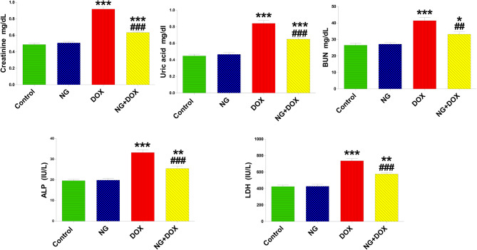
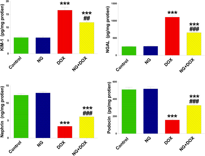
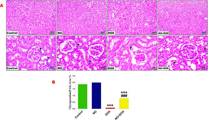
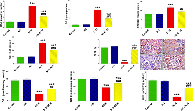
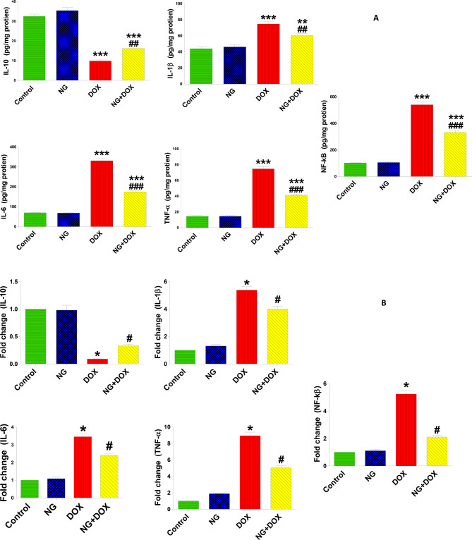
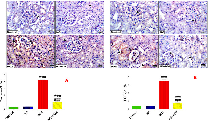
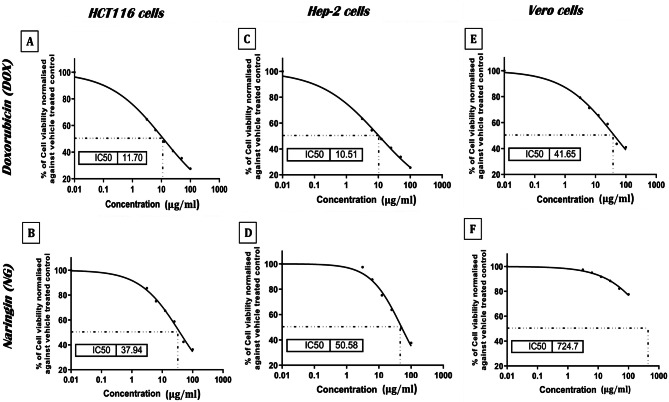
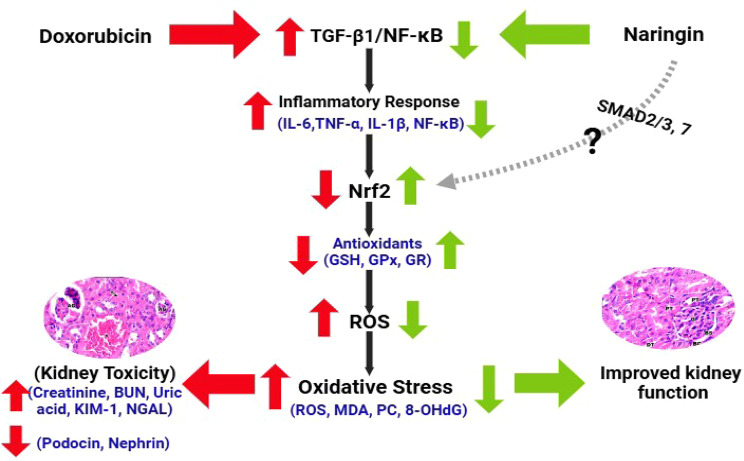
Results: Kidney damage and dysfunction were indicated by an elevation in the levels of creatinine, urea, uric acid, and the activity of ALP and LDH in serum, KIM-1, and NAGAL in kidney, and a significant decrease in nephrin and podocin in renal tissue. These disrupted glomerular and tubular function indicators were remarkably ameliorated by oral administration of NG (100 mg/kg) daily for 10 days before DOX treatment and continued for an additional four days post-Dox treatment. The nephroprotective effect of NG was confirmed by the improvement of histopathological and PAS histochemical investigations. The mitigating impact of NG was verified by normalization of the redox balance, evidenced by a significant amelioration of ROS levels, oxidative stress markers (MDA, PC, 8-OHdG), and antioxidants (GSH, GPx, GR), as well as upregulation of Nrf2 expression in kidney. Furthermore, NG significantly prevented the increase in the inflammatory mediators (IL-6, IL-1β, TNF-α, and NF-κB) and upregulated the anti-inflammatory IL-10 in DOX-treated rats. The expression of TGF-β1 and the apoptotic protein caspase-3 in the kidneys significantly decreased as a result of the improvement in redox state in renal tissue. Additionally, NG demonstrated anticancer effects and their combination showed synergistic anticancer impact on larynx and colon cancer cell lines in vitro study.

Conclusions: NG demonstrated remarkable protection of kidney against DOX treatment.

Keywords: Antioxidants; Doxorubicin; Inflammation; Kidney histopathology; Kidney injury; Naringin.


Conflict of interest statement

Declarations. Ethics approval and consent to participate: Institutional animal care and use committee (ACUC) of Mansoura University approved the legislation used in this work, which complies with international standards for laboratory animal handling and care (approval number: MU-ACUC (SC. PhD. 23.09.13)). Consent for publication: Not applicable Competing interests: The authors declare no competing interests.

Figures

Fig. 1
Fig. 1
Schematic representation of the experimental design. Rats were given 100 mg/kg of naringin (NG) orally for 14 days, and on the tenth day of the examination, they received an intraperitoneal (ip) injection of doxorubicin (DOX) at a dose of 15 mg/kg. After 14 days of the experiment, overnight fasted animals were euthanized then sacrificed, followed by samples collection
Fig. 2
Fig. 2
The effects of NG (100 mg/kg) and DOX (15 mg/kg) on the levels of creatinine, uric acid, blood urea nitrogen (BUN), alkaline phosphatase activity (ALP), and lactate dehydrogenase activity (LDH) in the serum of control and different treatment groups after 14 days. Values are expressed as mean ± SD (n = 5). *, ** and *** indicate statistical significance at P < 0.05, P < 0.01 and P < 0.001, respectively, compared to the control group. ## and ### indicate statistical significance at P < 0.01 and P < 0.001, respectively, compared to the DOX group
Fig. 3
Fig. 3
The effects of NG (100 mg/kg) and DOX (15 mg/kg) on the levels of kidney injury molecule-1 (KIM-1), neutrophil gelatinase-associated lipocalin (NGAL), nephrin, and podocin in the kidney tissues of the rats in the different groups. Values are expressed as mean ± SD (n = 5). *** indicate statistical significance at P < 0.001, compared to the control group. ## and ### indicate statistical significance at P < 0.01 and P < 0.001, respectively, compared to the DOX group
Fig. 4
Fig. 4
Photomicrographs of rat kidney sections from the control and different treatment groups (A). Control and NG groups revealing normal histological architecture represented by normal glomeruli (G), Bowman’s capsules (BC), and Bowman’s space (BS), as well as proximal convoluted tubule (PT) and distal convoluted tubule (DT). Sections of Dox-treated rats showing glomerular atrophy (AG), large and wide capsular space (arrow), dilation and congestion (*), as well as destruction of tubular cells and tubular necrosis (crossed arrow) and leukocytic infiltration (arrow head). Sections of NG + Dox-treated rats demonstrating retained normal histology of glomeruli and tubules. (H&E stain, X100&400). (B and C) Quantification is expressed as the perimeter of glomeruli and Bowman’s capsule (µm) in all studied groups, respectively. Values are expressed as the mean ± SD of 5 microscopic fields/tissue samples. * and *** indicate statistical significance at P < 0.05 and P < 0.001 respectively compared to the control group. ## indicate statistical significance at P < 0.01 compared to the DOX group
Fig. 5
Fig. 5
Periodic Acid Schiff (PAS) stained kidney sections of control and NG groups showing a strong positive reaction of polysaccharides in the glomerular area, Bowman’s capsule, and tubular area as well (head arrow) (A). Dox-treated rats revealed a significant (P < 0.001) reduction in the carbohydrate content of glomerular and many renal tubules (arrowhead). Sections of Dox + NG-treated rats exhibiting significant (P < 0.001) positive PAS reactions in glomerular and many renal tubules compared with those of the Dox-treated group (PAS X100 & 400). (B) Quantification of the PAS reaction in different treatment groups. Values are expressed as the means ± SD. *** indicate statistical significance at P < 0.001 compared to the control group. ### indicate statistical significance at P < 0.001 compared to the DOX group
Fig. 6
Fig. 6
The effects of NG (100 mg/kg) and DOX (15 mg/kg) on the levels of oxidative stress biomarkers: malondialdehyde (MDA), protein carbonyl (PC), 8-hydroxy-2’-deoxyguanosine (8-OHdG), and reactive oxygen species (ROS), as well as the antioxidants, including glutathione peroxidase (GPx) and glutathione reductase (GR) activities, and glutathione (GSH) content in kidney tissues of the different treatment groups. Data are expressed as mean ± SD (n = 5). Immunohistochemical expression of Nrf2 levels in the kidney sections of control and different animal groups. Control and NG groups displayed a strong expression of Nrf2 (arrowhead). Dox-treated animals showed weak Nrf2 immunoexpression (arrowhead). Dox + NG-treated rats revealed a marked Nrf2 expression to almost normal (arrowhead) (IHC, X400). Values are expressed as the means ± SD of 5 microscopic fields/tissue samples of Nrf2 immunoexpression. *** indicate statistical significance at P < 0.001, compared to the control group. ## and ### indicate statistical significance at P < 0.01 and P < 0.001, respectively, compared to the DOX group
Fig. 7
Fig. 7
The effects of NG (100 mg/kg) and DOX (15 mg/kg) on the levels of inflammatory cytokines. anti-inflammatory; interleukin 10 (IL-10) and pro-inflammatory; interleukin 1 beta (IL-1β), interleukin 6 (IL-6), Tumor necrosis factor alpha (TNFα), and nuclear factor kappa B (NF-κB) in the kidneys of the rats in the different groups. Values are expressed as mean ± SD (n = 5). Figure 7 B. RT-qPCR analysis of (IL-10), (IL-1β), (IL-6), (TNFα), and (NF–kB) in relative gene expression kidney tissues in the different groups. Results are expressed as mean (fold change) ± SD (n = 5). ** and *** indicate statistical significance at P < 0.01 and P < 0.001, respectively, compared to the control group. ## and ### indicate statistical significance at P < 0.01 and P < 0.001, respectively, compared to the DOX group
Fig. 8
Fig. 8
Immunohistochemical determination of the expression of caspase-3 and TGF-β1 in the kidney sections of control and different animal groups (A). Expression indicated by arrowhead (IHC, X400). (B) Values are expressed as the means ± SD of 5 microscopic fields/tissue samples of caspase-3 and TGF-β1 immunoexpression. ###, ***Very highly significant at P < 0.001. *** Significant as compared with the control group. ### Significant as compared with the Dox-treated group. *** indicate statistical significance at P < 0.001, compared to the control group. ### indicate statistical significance at P < 0.001, compared to the DOX group
Fig. 9
Fig. 9
Sigmoidal curve for MTT assay showing IC50 values and the inhibition % of DOX and NG on HCT116, Hep-2 and Vero cells after 24 hrs treatment. Each data point represents an average of three independent experiments (n = 5)
Fig. 10
Fig. 10
Putative mechanisms of the TGF-β1, NF-κB/Nrf2 signaling crosstalk in DOX-induced kidney injury. Under kidney lesions induced by DOX treatment, TGF-β1 release increases, most likely by podocyte and kidney cells. The activation of the TGF-β1 will, in turn, deactivate the Nrf2-dependent antioxidant response, including the inhibition of antioxidant enzymes and the development of oxidative stress. Thus, blocking the TGF-β1 receptor and its release by NG reduces this response through mechanisms that are yet to be identified (?), triggering a failure in antioxidant-related recovery

Similar articles

Cited by

References

    1. Ibrahim Fouad G, Ahmed KA. The protective impact of berberine against doxorubicin-induced nephrotoxicity in rats. Tissue Cell. 2021;73:101612. - PubMed
    1. Al-Naimi MS, et al. Nephrotoxicity: role and significance of renal biomarkers in the early detection of acute renal injury. J Adv Pharm Technol Res. 2019;10:95–99. - PMC - PubMed
    1. Afsar T, et al. Doxorubicin-induced alterations in kidney functioning, oxidative stress, DNA damage, and renal tissue morphology; Improvement by Acacia hydaspica tannin-rich ethyl acetate fraction. Saudi J Biol Sci. 2020;27:2251–60. - PMC - PubMed
    1. Sang Y, et al. Semaphorin3A-inhibitor ameliorates doxorubicin-induced podocyte injury. Int J Mol Sci. 2020;21(11). - PMC - PubMed
    1. Pan QR, et al. Resveratrol increases nephrin and podocin expression and alleviates renal damage in rats fed a high-fat diet. Nutrients. 2014;6:2619–31. - PMC - PubMed

MeSH terms