. 2025 Jun 23;11(3):00732-2024.
doi: 10.1183/23120541.00732-2024. eCollection 2025 May.

Lung parenchymal and cardiac appearances on computed tomography pulmonary angiography impact survival in chronic thromboembolic pulmonary hypertension: results from the ASPIRE Registry

Affiliations

Lung parenchymal and cardiac appearances on computed tomography pulmonary angiography impact survival in chronic thromboembolic pulmonary hypertension: results from the ASPIRE Registry

Lojain Abdulaal et al. ERJ Open Res. .

Abstract

Background: Chronic thromboembolic pulmonary hypertension (CTEPH) is commonly evaluated using computed tomography pulmonary angiography (CTPA). We evaluated the frequency and impact of parenchymal and cardiac abnormalities on survival in CTEPH.

Methods: Patients were identified from the ASPIRE (Assessing the Spectrum of Pulmonary Hypertension Identified at a Referral Centre) Registry. Kaplan-Meier analysis was used to assess survival.

Results: 290 patients (55% female, mean±sd age 65±14 years) with CTEPH were included. Mosaic perfusion was noted in 83%, lung infarction in 73% and parenchymal lung disease in 28%. The severity of mosaic perfusion and lung infarction correlated with markers of disease severity (p<0.001). Whereas the presence of mosaic perfusion was associated with improved survival in all patients (p=0.03), it did not predict outcome in those undergoing pulmonary endarterectomy (PEA) (p=0.6) and those not undergoing PEA (p=0.22). The presence of lung infarction had no impact on mortality. The presence of co-existing lung disease was associated with a worse survival (p<0.008) in patients not undergoing PEA. Mosaic perfusion was less common in patients with parenchymal lung disease (65%) compared to those without parenchymal lung disease (90%), p<0.001. An increased right/left ventricular ratio and aortic diameter predicted a worse outcome (p<0.002).

Conclusion: Lung parenchymal and cardiac changes on CTPA predict outcome in CTEPH. Co-existing parenchymal lung disease is not uncommon and when present may mask the presence of mosaic perfusion. This study highlights the importance of systematically evaluating the lung parenchyma and cardiac changes in patients with CTEPH.


Conflict of interest statement

Conflict of interest: D.G. Kiely, S. Alabed and A.J. Swift are investigators within the National Institute for Health Research (NIHR) Sheffield Cardiovascular Biomedical Research Unit. A.J. Swift reports research grants and speaker fees from Janssen, the Wellcome Trust and the BHF. M. Sharkey is funded by the Wellcome Trust (223521/Z/21/Z) and NIHR Sheffield Biomedical Research Centre (NIHR203321). The authors declare that the research was conducted in the absence of any commercial or financial relationships that could be construed as a potential conflict of interest.

Figures
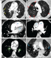
FIGURE 1
FIGURE 1
a) Examples of lung parenchymal features of chronic thromboembolic pulmonary hypertension (CTEPH) on computed tomography pulmonary angiography (CTPA). Axial CTPA images with standard lung windows from two different patients. The patient on the left showed lung infarction within the right middle lobe (yellow arrows). This typically appears as focal wedge-shaped consolidation at the lung periphery which resolves over time. Both patients exhibit mosaic perfusion. This refers to regional hyperattenuation (dashed red arrows) and hypoattenuation (solid red arrows) of the lung parenchyma, of which abnormal lung perfusion is a cause. It is important to note that neither lung infarction nor mosaic perfusion are specific to CTEPH and there is a broad differential diagnosis for both of these imaging findings. b) Examples of cardiac changes commonly seen in CTEPH on CTPA. Two axial CTPA images with standard soft tissue windows are shown. The right image shows marked dilatation of the right ventricle (RV) relative to the left ventricle (LV), with an RV/LV ratio of > 1.0, consistent with RV dysfunction. The left image shows dilatation of the pulmonary artery (PA) trunk relative to the ascending aorta (AA), with a PA/AA ratio of >1.0. c) Examples of CTEPH within left and right segmental disease (green circles), and emphysema lung disease in both lungs (blue arrows).
FIGURE 2
FIGURE 2
Study flow diagram. CT: computed tomography; ASPIRE: Assessing the Spectrum of Pulmonary Hypertension Identified at a Referral Centre; PH: pulmonary hypertension; CTEPH: chronic thromboembolic pulmonary hypertension; CTPA: computed tomography pulmonary angiography.
FIGURE 3
FIGURE 3
Group comparison of the extent of lung mosaic perfusion (top) and lung infarction (bottom) with right heart catheterisation and TLCO using one-way ANOVA. Pulmonary vascular resistance (PVR), mean pulmonary arterial pressure (mPAP), venous oxygen saturation (SvO2), and transfer factor of the lung for carbon monoxide (TLCO). NS: nonsignificant.
FIGURE 4
FIGURE 4
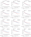
First Kaplan–Meier survival curve comparing chronic thromboembolic pulmonary hypertension patients who underwent pulmonary endarterectomy (PEA) to those who did not. along with additional curves predicting mosaic perfusion, infarction and lung disease related mortality for patients who had PEA and who did not undergo PEA after computed tomography (CT). These curves are based on CT scan findings and show numbers at risk by years.
FIGURE 5
FIGURE 5
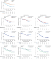
Kaplan–Meier survival curves examining the impact of computed tomography pulmonary angiography measured vessels and cardiac chambers on survival in all patients with chronic thromboembolic pulmonary hypertension, those undergoing pulmonary endarterectomy (PEA) and those not undergoing PEA (pulmonary artery (PA), aorta (AO), PA/AO ratio and right ventricle (RV)/left ventricle (LV) ratio).

Similar articles

References

    1. Delcroix M, Torbicki A, Gopalan D, et al. ERS statement on chronic thromboembolic pulmonary hypertension. Eur Respir J 2021; 57: 2002828. doi: 10.1183/13993003.02828-2020 - DOI - PubMed
    1. Valerio L, Mavromanoli AC, Barco S, et al. Chronic thromboembolic pulmonary hypertension and impairment after pulmonary embolism: the FOCUS study. Eur Heart J 2022; 43: 3387–3398. doi: 10.1093/eurheartj/ehac206 - DOI - PMC - PubMed
    1. Durrington C, Hurdman JA, Elliot CA, et al. Systematic pulmonary embolism follow-up increases diagnostic rates of chronic thromboembolic pulmonary hypertension and identifies less severe disease: results from the ASPIRE Registry. Eur Respir J 2024; 63: 2300846. doi: 10.1183/13993003.00846-2023 - DOI - PMC - PubMed
    1. Pepke-Zaba J, Delcroix M, Lang I, et al. Chronic thromboembolic pulmonary hypertension (CTEPH) results from an international prospective registry. Circulation 2011; 124: 1973–1981. doi: 10.1161/CIRCULATIONAHA.110.015008 - DOI - PubMed
    1. Quadery SR, Swift AJ, Billings CG, et al. The impact of patient choice on survival in chronic thromboembolic pulmonary hypertension. Eur Respir J 2018; 52: 1800589. doi: 10.1183/13993003.00589-2018 - DOI - PMC - PubMed

LinkOut - more resources