Acute Distal Internal Carotid Artery Occlusion in Which Angiography during Mechanical Thrombectomy Revealed a Shunt between the Internal Carotid Artery and the Cavernous Sinus: A Case Report
- PMID: 40551878
- PMCID: PMC12185224
- DOI: 10.5797/jnet.cr.2025-0033
Acute Distal Internal Carotid Artery Occlusion in Which Angiography during Mechanical Thrombectomy Revealed a Shunt between the Internal Carotid Artery and the Cavernous Sinus: A Case Report
Abstract
Objective: We report a patient with occlusion of the distal internal carotid artery (ICA), in whom angiography during mechanical thrombectomy revealed a shunt between the ICA and the cavernous sinus.
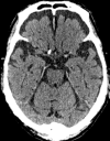
Case presentation: A 79-year-old man with bile duct cancer, a liver abscess, septic shock, and atrial fibrillation presented to our hospital with sudden disturbance of consciousness, conjugate eye deviation, and right hemiplegia. A cranial CT revealed a hyperdense middle cerebral artery (MCA) and loss of gray-white matter differentiation, suggesting large vessel occlusion. Endovascular therapy was immediately initiated. Left internal carotid angiography indicated occlusion of the distal ICA at the origin of the ophthalmic artery. Injection of contrast medium at a site just proximal to the ICA occlusion depicted the cavernous sinus and inferior petrosal sinus. We withdrew the aspiration catheter to the petrous segment of the ICA and injected contrast medium again. This time, however, neither the cavernous sinus nor the inferior petrosal sinus was visualized. We deployed a stent retriever at the occlusion site and successfully removed the thrombus. The final angiography showed complete recanalization of the affected arterial segment with no sign of a carotid cavernous fistula. The patient was finally discharged on day 73 after endovascular therapy with a cerebral infarction in the territory of the left MCA.
Conclusion: In the present case, angiographic visualization of the cavernous sinus varied depending on the site of contrast medium injection. It appears that the high pressure of the contrast medium generated in the stump of the ICA opened up microvascular shunts between the normal capillaries of the ICA and the cavernous sinus, leading to visualization of the cavernous sinus. Therefore, it is important to be aware that injection of contrast medium into the blind alley of the ICA near the cavernous sinus could result in early visualization of the cavernous sinus.
Keywords: cavernous sinus; large vessel occlusion; mechanical thrombectomy.
©2025 The Japanese Society for Neuroendovascular Therapy.
Figures


Similar articles
-
Safe Endovascular Recanalization of Internal Carotid Artery Occlusion Using Retrograde Aspiration Angiography.World Neurosurg. 2024 Dec;192:162-169. doi: 10.1016/j.wneu.2024.09.133. Epub 2024 Oct 22. World Neurosurg. 2024. PMID: 39366480
-
Urgent carotid endarterectomy with distal mechanical thrombectomy.J Neurointerv Surg. 2025 Jan 25:jnis-2024-021662. doi: 10.1136/jnis-2024-021662. Online ahead of print. J Neurointerv Surg. 2025. PMID: 38906686
-
Thrombectomy using Zoom Reperfusion System in pediatric acute ischemic strokes: Case series and literature review.Neuroradiol J. 2025 Jun;38(3):349-353. doi: 10.1177/19714009241247468. Epub 2024 May 2. Neuroradiol J. 2025. PMID: 38695294 Free PMC article. Review.
-
Ruptured Primitive Trigeminal Artery Aneurysm Leading to Internal Carotid Artery Cavernous Sinus Fistula Intervention.J Craniofac Surg. 2025 Sep 1;36(6):e614-e616. doi: 10.1097/SCS.0000000000011098. Epub 2025 Feb 11. J Craniofac Surg. 2025. PMID: 39932795
-
Duplex ultrasound for diagnosing symptomatic carotid stenosis in the extracranial segments.Cochrane Database Syst Rev. 2022 Jul 11;7(7):CD013172. doi: 10.1002/14651858.CD013172.pub2. Cochrane Database Syst Rev. 2022. PMID: 35815652 Free PMC article.
References
-
- Sarraj A, Hassan AE, Abraham MG, et al. Trial of endovascular thrombectomy for large ischemic strokes. N Engl J Med 2023; 388: 1259–1271. - PubMed
-
- Grüter BE, Kahles T, Anon J, et al. Carotid-cavernous sinus fistula following mechanical thrombectomy in acute ischaemic stroke: a rare complication. Neuroradiology 2021; 63: 1149–1152. - PubMed
-
- Alan N, Nwachuku E, Jovin TJ, et al. Management of iatrogenic direct carotid cavernous fistula occurring during endovascular treatment of stroke. World Neurosurg 2017; 100: 710.e15–710.e20. - PubMed
Publication types
LinkOut - more resources
Full Text Sources
Miscellaneous

