

[Buffalo Horn Sign - A New Finding on MRI for Meniscal Bucket-Handle Tears]
- PMID: 40552002
- PMCID: PMC12185184
- DOI: 10.1055/s-0045-1809514
[Buffalo Horn Sign - A New Finding on MRI for Meniscal Bucket-Handle Tears]
Abstract
Objective: To describe a new sign on magnetic resonance imaging (MRI) axial images of patients with bucket-handle meniscal tears.
Methods: Of 610 consecutive patients with a surgical diagnosis of meniscal tear, those with a bucket-handle pattern were chosen, and 28 met the inclusion criteria. The most frequent mechanism was a twisting injury with or without a coronal stress (16 patients), and the injury was sports-related in 12 cases. All patients were symptomatic and had X-rays showing a preserved joint line. Next, their MRI examinations were analyzed.
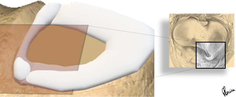
Results: The buffalo horn pattern was found in 13 patients (46.4%), occurring in either the medial or the lateral meniscus. It was the 3 rd most prevalent sign, after the fragment within the intercondylar notch ( n = 21; 75.0%) and the absent bow tie sign ( n = 17; 60.7%). We observed that it had a significant association with other signs of displaced meniscal handle. The sign was neither found on the healthy menisci, nor was affected by the occurrence of an anterior cruciate ligament tear.
Conclusion: The buffalo horn is a new finding for displaced meniscal bucket-handle tears; it is easy to identify and relevant in the interpretation of axial MRI images. Its recognition is very important to determine the type of treatment and the surgical plan.
Objetivo: Descrever um novo sinal em imagens axiais de ressonância magnética (RM) de pacientes com rupturas em alça de balde do menisco.
Métodos: De 610 pacientes consecutivos com diagnóstico cirúrgico de ruptura do menisco, aqueles com padrão em alça de balde foram escolhidos, e 28 atenderam aos critérios de inclusão. O mecanismo de lesão mais frequente foi a torção com ou sem estresse coronal (16 pacientes). Além disso, a lesão foi relacionada ao esporte em 12 casos. Todos os pacientes eram sintomáticos e tinham radiografias que mostravam a preservação da linha articular. Em seguida, seus exames de RM foram analisados.
Resultados: O padrão de chifre de búfalo foi encontrado em 13 pacientes (46,4%) no menisco medial ou lateral. Foi o 3 o sinal mais prevalente, depois do fragmento no interior da incisura intercondilar ( n = 21; 75,0%) e da ausência do sinal da gravata borboleta ( n = 17; 60,7%). Observamos uma associação significativa a outros sinais de deslocamento da alça do menisco. O sinal não foi encontrado em meniscos saudáveis, nem foi afetado pela ocorrência de ruptura do ligamento cruzado anterior.
Conclusão: O chifre de búfalo é um novo achado para rupturas em alça de balde do menisco com deslocamento; é fácil de identificar e relevante na interpretação de imagens de RM de corte axial. Seu reconhecimento é muito importante para determinar o tipo de tratamento e o plano cirúrgico.
Keywords: diagnostic imaging; knee; magnetic resonance imaging; tibial meniscus injuries.
The Author(s). This is an open access article published by Thieme under the terms of the Creative Commons Attribution 4.0 International License, permitting copying and reproduction so long as the original work is given appropriate credit ( https://creativecommons.org/licenses/by/4.0/ ).
Conflict of interest statement
Conflito de Interesses Os autores não têm conflito de interesses a declarar.
Figures


References
Publication types
LinkOut - more resources
Full Text Sources
Miscellaneous