. 2025 Jun 9:250:10598.
doi: 10.3389/ebm.2025.10598. eCollection 2025.

Cystamine reduces neurodegeneration and epileptogenesis following soman-induced status epilepticus in rats

Affiliations

Cystamine reduces neurodegeneration and epileptogenesis following soman-induced status epilepticus in rats

Abiel K Biney et al. Exp Biol Med (Maywood). .

Abstract

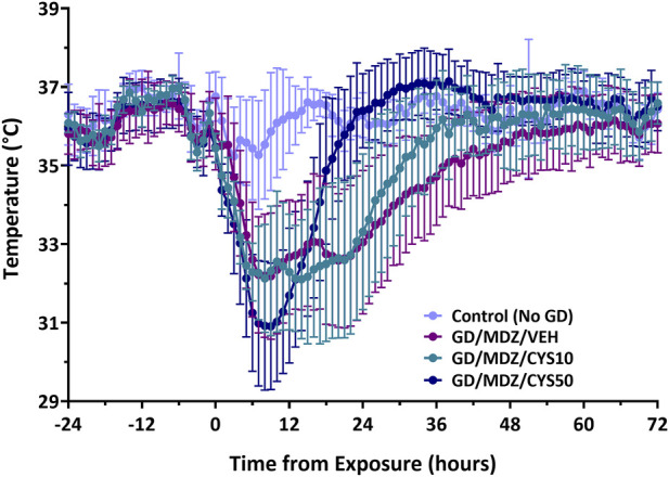
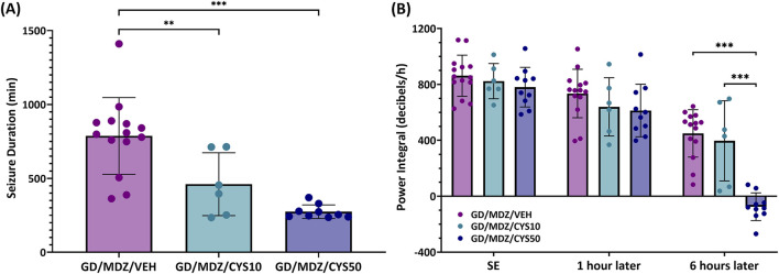
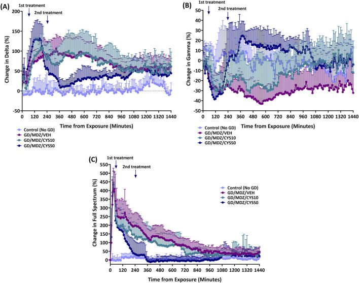
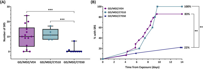
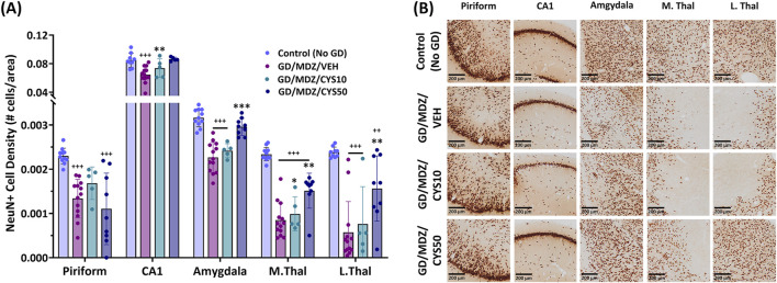
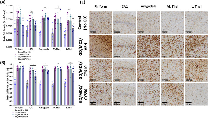
Acute exposure to a seizure-inducing dose of an organophosphorus nerve agent inhibits acetylcholinesterase, leading to pharmacoresistance if benzodiazepine treatment is delayed. Following soman-induced status epilepticus (SE) in rats, prolonged seizure is associated with severe and widespread neurodegeneration. We evaluated the aminothiol cystamine, the oxidized form of cysteamine, for neuroprotective potential against soman-induced SE and associated neurodegeneration. Cystamine has a myriad of effects including antioxidant properties, neuroprotective effects, and immunomodulation, among others, which is of interest in evaluating neuroprotective efficacy against cholinergic-induced neurodegeneration. Adult male rats implanted with telemetry transmitters for continuous EEG recording were exposed to soman and treated with the muscarinic antagonist atropine sulfate and the oxime asoxime dimethanesulfonate 1 min after exposure to increase survival. Midazolam was administered 30 min after seizure onset. Cystamine (10 or 50 mg/kg) or vehicle was administered 30 min after seizure onset and again 4 h after soman exposure. The initial seizure duration, the EEG power integral at 6 h after exposure, and the percentage of rats that developed spontaneous recurrent seizure were reduced in rats treated with cystamine, compared to those that received only midazolam. In addition, cystamine reduced neurodegeneration in seizure-sensitive brain regions following soman exposure, compared to midazolam. Our findings highlight the potential for aminothiols to serve as adjunctive therapy to midazolam in treating cholinergic-induced toxicity and suggest broader applications of aminothiols in neuroprotection and neurological disorders.

Keywords: cystamine; neuroprotection; organophosphorus nerve agents; seizures; status epilepticus.


Conflict of interest statement

Author MA was employed by BioSEaD, LLC. The remaining author declared no potential conflicts of interest with respect to the research, authorship, and/or publication of this article.

Figures

FIGURE 1
FIGURE 1
The effect of cystamine treatment on body temperature in adult male rats exposed to soman. Rats exposed to soman were treated with atropine sulfate and HI-6 1 minute after exposure and with midazolam (GD/MDZ/VEH) with or without 10 mg/kg cystamine (GD/MDZ/CYS10) or 50 mg/kg cystamine (GD/MDZ/CYS50) 30 min and 4 h after seizure onset. In all soman-exposed groups, body temperature was reduced within a few hours compared to baseline and to Control (No GD). The GD/MDZ/CYS50 group had lower body temperature at 1 h and 2 h compared to the GD/MDZ/VEH group (p < 0.05), yet recovered body temperature more rapidly; from 18 to 44 h, 46–48 h, and 56 h after exposure. The GD/MDZ/CYS50 rats had significantly higher body temperature compared to GD/MDZ/VEH (p < 0.05). In addition, the GD/MDZ/CYS50 group returned to baseline temperature by 18 h, the GD/MDZ/CYS10 within 26 h and the GD/MDZ/VEH within 37 h after exposure. n = 11 for Control (No GD), n = 14 for GD/MDZ/VEH, n = 6 for GD/MDZ/CYS10, n = 9 for GD/MDZ/CYS50. Data are shown as mean ± SD.
FIGURE 2
FIGURE 2
The effect of cystamine treatment on initial seizure duration and power integral in adult male rats exposed to soman. Rats exposed to soman were treated with atropine sulfate and HI-6 1 minute after exposure and with midazolam (GD/MDZ/VEH) with or without 10 mg/kg cystamine (GD/MDZ/CYS10) or 50 mg/kg cystamine (GD/MDZ/CYS50) 30 min and 4 h after seizure onset. (A) Adult male rats exposed to soman and treated with the cystamine (10 or 50 mg/kg) had reduced duration of initial seizures during the first 24 h compared to midazolam-treated rats (***p < 0.001; **p < 0.01). n = 14 for GD/MDZ/VEH, n = 6 for GD/MDZ/CYS10, n = 9 for GD/MDZ/CYS50. (B) Comparison of power integral at SE onset, 1 h after treatment, and 6 h after treatment. Power integral is averaged over 10 min time bins. Of the cystamine groups, neither showed differences from midazolam monotherapy at 1 h. Only the 50 mg/kg dose showed a significant reduction in the EEG power integral at the 6 h post-treatment period compared to midazolam monotherapy and compared to the lower cystamine dose (***p < 0.001). n = 14 for GD/MDZ/VEH, n = 6 for GD/MDZ/CYS10, n = 10 for GD/MDZ/CYS50. Data are shown as mean ± SD.
FIGURE 3
FIGURE 3
The percent of relative change from baseline levels in the power of (A) delta band (0.1–4 Hz), (B) gamma band (25.1–50 Hz), and (C) total power spectra was calculated for soman-exposed rats that received midazolam (GD/MDZ/VEH) or cystamine as adjunct to midazolam (GD/MDZ/CYS10; GD/MDZ/CYS50). The GD/MDZ/CYS50 group had reduced change in delta band activity, increased change in gamma band, and reduced change in total EEG power spectra compared to soman-exposed rats that received midazolam (GD/MDZ/VEH). (A) For delta p < 0.05 in comparison of Control (No GD) vs. GD/MDZ/CYS50 at 10–220, 250, 290, 600–610, 650, 720–770, 800–1000, 1020–1200, 1230, and 1380–1440 min after exposure; GD/MDZ/VEH vs. GD/MDZ/CYS50 at 20, 50, 110–170, 280–880, 960–970, and 990–1000 min after exposure. (B) For gamma p < 0.05 in comparison of Control (No GD) vs. GD/MDZ/CYS50 at 10–20, 40–190, 400, 460–480, 500, 680–690, 790, and 850–870 min after exposure; GD/MDZ/VEH vs. GD/MDZ/CYS50 at 260–1070, 1140–1150, 1210, 1230, 1260–1270, 1300, 1320, 1340–1350, 1370, 1400, 1420, and 1440 min after exposure. (C) For full spectrum p < 0.05 in comparison of Control (No GD) vs. GD/MDZ/CYS50 at 10–130 and 150–160 min after exposure; GD/MDZ/VEH vs. GD/MDZ/CYS50 at 140–1070 min after exposure. n = 11 for Control (No GD), n = 14 for GD/MDZ/VEH, n = 6 for GD/MDZ/CYS10, n = 9 for GD/MDZ/CYS50.
FIGURE 4
FIGURE 4
The effect of cystamine on the development of spontaneous recurrent seizures (SRS) following soman exposure in adult male rats. Rats exposed to soman were treated with atropine sulfate and HI-6 1 minute after exposure and with midazolam (GD/MDZ/VEH) with or without 10 mg/kg cystamine (GD/MDZ/CYS/10) or 50 mg/kg cystamine (GD/MDZ/CYS50) 30 min and 4 h after seizure onset. (A) Rats in the GD/MDZ/CYS50 group had fewer SRS occurrences compared to the GD/MDZ/VEH group and GD/MDZ/CYS10 group (***p < 0.001). n = 14 for GD/MDZ/VEH, n = 5 for GD/MDZ/CYS10, n = 9 for GD/MDZ/CYS50. (B) Onset of epileptogenesis is shown over a 14-day period from exposure. An effect of group was found on the median onset of SRS when comparing the GD/MDZ/CYS50 group to the GD/MDZ/VEH group (p < 0.01). The GD/MDZ/CYS50 group had a smaller percentage of rats that developed SRS compared to GD/MDZ/VEH and to GD/MDZ/CYS50. %SRS (**p < 0.01 at final % of animals with SRS). n = 14 for GD/MDZ/VEH, n = 5 for GD/MDZ/CYS10, n = 9 for GD/MDZ/CYS50.
FIGURE 5
FIGURE 5
Soman exposure in adult male rats induced widespread neuronal loss in the piriform cortex, CA1 of the hippocampus, basolateral amygdala, medial thalamus (M. Thal), and lateral thalamus (L. Thal), shown here 2 weeks after exposure. Comparisons are made between Control (No GD), midazolam treatment (GD/MDZ/VEH), and combination midazolam and cystamine treatment groups (GD/MDZ/CYS10; GD/MDZ/CYS50). (A) Rats treated with the 50 mg/kg cystamine dose adjunct with midazolam had reduced neuronal loss in the CA1, amygdala, M. Thal, and L. Thal compared to midazolam monotherapy. Compared to GD/MDZ/VEH: **p < 0.01; ***p < 0.001. Compared to Control (No GD): ++p < 0.01; +++p < 0.001. n = 11 for Control (No GD), n = 14 for GD/MDZ/VEH, n = 5 for GD/MDZ/CYS10, n = 9 for GD/MDZ/CYS50. Data are shown as mean ± SD. (B) Representative photomicrographs of NeuN-stained coronal sections are shown at the 14-day endpoint depicting the piriform, CA1, amygdala, M. Thal, and L. Thal. Images are taken at ×10 magnification (scale bar = 200 µm).
FIGURE 6
FIGURE 6
Soman exposure in adult male rats induced a severe neuroinflammatory response in the piriform cortex, CA1 of the hippocampus, basolateral amygdala, medial thalamus (M.Thal), and lateral thalamus (L.Thal), demonstrated with Iba+-staining. Comparisons are made between Control (No GD), midazolam (GD/MDZ/VEH), and combination midazolam and cystamine treatment groups (GD/MDZ/CYS10; GD/MDZ/CYS50). The GD/MDZ/CYS50 group (A) attenuated soman-induced increase in microglia density in the L. Thal and (B) mitigated microglial activation in the CA1 compared to the GD/MDZ/VEH group (**p < 0.01). The 10 mg/kg dose did not demonstrate significant protection against neuroinflammatory responses. Compared to GD/MDZ/VEH: **p < 0.01; Compared to Control (No GD): +p < 0.05; +++p < 0.001. n = 11 for Control (No GD), n = 14 for GD/MDZ, n = 5 for GD/MDZ/CYS10, n = 9 for GD/MDZ/CYS50. Data are shown as mean ± SD. (C) Representative photomicrographs of Iba1-stained coronal sections with a cresyl violet counterstain are shown at the 14-day endpoint depicting the piriform, CA1, amygdala, M. Thal, and L. Thal. Images were taken at ×20 magnification (scale bar = 100 µm).

Similar articles

Cited by

References

    1. Paul BD, Snyder SH. Therapeutic applications of cysteamine and cystamine in neurodegenerative and neuropsychiatric diseases. Front Neurol (2019) 10:1315. 10.3389/fneur.2019.01315 - DOI - PMC - PubMed
    1. Borrell-Pages M, Canals JM, Cordelieres FP, Parker JA, Pineda JR, Grange G, et al. Cystamine and cysteamine increase brain levels of BDNF in Huntington disease via HSJ1b and transglutaminase. J Clin Invest (2006) 116:1410–24. 10.1172/JCI27607 - DOI - PMC - PubMed
    1. Bousquet M, Gibrat C, Ouellet M, Rouillard C, Calon F, Cicchetti F. Cystamine metabolism and brain transport properties: clinical implications for neurodegenerative diseases. J Neurochem (2010) 114:1651–8. 10.1111/j.1471-4159.2010.06874.x - DOI - PubMed
    1. Arbez N, Roby E, Akimov S, Eddings C, Ren M, Wang X, et al. Cysteamine protects neurons from mutant huntingtin toxicity. J Huntington's Dis (2019) 8:129–43. 10.3233/jhd-180312 - DOI - PMC - PubMed
    1. Gibrat C, Bousquet M, Saint-Pierre M, Levesque D, Calon F, Rouillard C, et al. Cystamine prevents MPTP-induced toxicity in young adult mice via the up-regulation of the brain-derived neurotrophic factor. Prog Neuro-Psychopharmacology Biol Psychiatry (2010) 34:193–203. 10.1016/j.pnpbp.2009.11.005 - DOI - PubMed

LinkOut - more resources