. 2024 Oct 13;1(4):100048.
doi: 10.1016/j.bneo.2024.100048. eCollection 2024 Dec.

Development and characterization of a low-affinity humanized CD19 chimeric antigen receptor for B-cell malignancies

Affiliations

Development and characterization of a low-affinity humanized CD19 chimeric antigen receptor for B-cell malignancies

Lawrence A Stern et al. Blood Neoplasia. .

Abstract

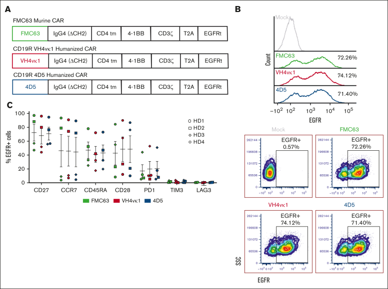
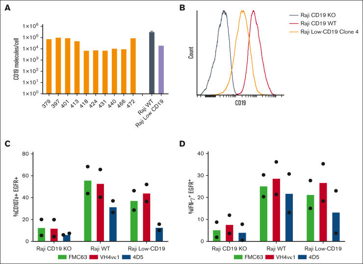
In this study, we aim to develop a humanized CD19 chimeric antigen receptor (CAR) that matches the potency of the FMC63 CAR and potentially reduces the risk of immunogenicity. The murine FMC63 single-chain variable fragment (scFv) was humanized yielding 2 lead candidate scFvs, VH4vκ1 and 4D5, which exhibit weaker binding affinity than FMC63 scFv. These humanized CD19-scFvs were incorporated into CAR constructs to generate huCD19R(VH4Vκ1) and huCD19R(4D5) CARs, both containing the 41BB costimulatory domain. The antitumor activity of the CAR T cells was assessed against CD19+ and CD19 low-expressing tumors. FMC63 CAR T cells with the same backbone in all studies were used as controls. The results showed that the huCD19R(VH4vκ1) CAR T cells exhibited similar expansion, phenotype, and effector function to the FMC63 CAR upon stimulation with CD19 targets. When the CAR T cells were challenged with CD19-bearing tumors, the huCD19R(VH4vκ1) CAR T cells showed similar proliferation to the FMC63 CAR T cells, whereas the huCD19R(4D5) CAR T cells essentially failed to proliferate. Moreover, the huCD19R(VH4vκ1) CAR T cells exhibited significantly better in vivo antitumor activity than the huCD19R(4D5) CAR T cells when tested against tumors expressing a range of CD19 antigens. Finally, using a hybrid model, we found that the huCD19R(VH4vκ1) T cells had a comparable cytokine secretion profile to that of FMC63 CAR T cells. Furthermore, the huCD19R(VH4vκ1) CAR T cells exhibited efficacy against both CD19+ and engineered CD19 low-expressing tumors. These findings suggest that huCD19R(VH4vκ1) CAR T cells may offer enhanced persistence and represent a promising candidate for clinical translation as a therapy for CD19+ tumors.


Conflict of interest statement

Conflict-of-interest disclosure: L.A.S., J.C.W., S.J.F. X.W., C.E.B. are the inventors of the huCD19CAR patent application. The remaining authors declare no competing financial interests. The current affiliation for L.A.S. is the Department of Chemical, Biological, and Materials Engineering, University of South Florida, Tampa, FL

Figures

None
Graphical abstract
Figure 1.
Figure 1.
Engineering and characterization of humanized CD19-targeted scFvs. (A) The murine anti-CD19 scFv FMC63 (black) was subjected to CDR grafting and in silico assessment, resulting in the generation of 2 humanized variants, huCD19R(4D5) (blue) and huCD19R(VH4vκ1) (red). (B) CD19-targeted scFvs (red with yellow CDRs) were displayed on the surface of yeast cells as N-terminal fusions to a flexible polypeptide linker (black) and agglutinin 2 (Aga2p; light blue). Fusions were anchored to the yeast cell wall via disulfide linkage to Aga1p (dark blue). scFv expression is detected by antibody labeling of a C-terminal c-Myc epitope (green) and antigen binding is detected using fluorescently tagged CD19 extracellular domain (purple). (C) Successful expression of FMC63 or humanized variants scFvs on the yeast surface was detected by c-Myc epitope tag labeling. One representative trial is shown. (D) The binding affinity of the indicated scFvs was tested using flow cytometry with titration of biotinylated recombinant CD19 extracellular domain and fit to a 1:1 equilibrium binding model. Data are presented as mean ± standard deviation of 3 trials. Titration curve fits are calculated based on the average estimated Kd for each clone. Concentrations on the x-axis are in nanomolar (nM). (E) The thermal stabilities of the indicated scFvs were determined by heating yeast displaying the indicated scFvs followed by labeling with either 50 nM biotinylated recombinant CD19 extracellular domain (FMC63) or conformation-specific binder biotinylated protein L (humanized variants). The fluorescence proportion of foldedness was determined by flow cytometry. Data are fit to a 2-state unfolding model. Thermal stabilities are presented as mean ± standard deviation of 3 to 4 trials.
Figure 2.
Figure 2.
CAR T-cell design and characterization. (A) Diagram of lentiviral cassette design for CD19-targeted CARs including CD19 scFv (FMC63, VH4vκ1, and 4D5), immunoglobulin G4 hinge, and CH3 connected by a 10-amino-acid glycine and serine (GS) linker (ch2Δ), CD4 transmembrane domain, cytoplasmic 4-1BB costimulatory domain, and cytoplasmic CD3ζ stimulatory domain. A non-signaling truncated EGFR is separated from the CAR by a T2A ribosome skip sequence for tracking transduction efficiency. (B) CD19 CARs were expressed in the healthy donor (HD) Tn/mem cells by lentiviral transduction. CAR expression is measured as % EGFR+. Representative histograms (top) and dot plots (bottom) are shown (n = 3). (C) Murine and humanized CAR T cells were characterized by analyzing the expressions of CD27, CCR7, CD45RA, CD28, programmed cell death protein 1 (PD1), TIM3, and LAG3 (n = 4). Data are shown as mean ± standard error of the mean (SEM).
Figure 3.
Figure 3.
Humanized CAR T cells exhibited antigen-specific effector function. (A) The growth curve for Mock, FMC63, VH4vκ1, and 4D5 is shown as fold change normalized to day 0. (B) Surface expression of CD19 antigen, costimulatory molecules CD80, CD86, and inhibitory ligand PD-L1 on tumor lines KG1a, Nalm6, and Raji. Tn/mem cells expressing the specified CAR constructs were analyzed for their ability to (C) degranulate (107a), (D) produce IFN-γ and in response to KG1a, CD19+ Raji, and CD19+ Nalm6 by flow cytometry. Data are shown as mean ± SEM for CAR+ CD107a+ (n = 4) and CAR+ IFN-γ (n = 3). Tn/mem cells expressing the specified CAR constructs were challenged with CD19+ green fluorescent protein-positive (GFP+) Raji cells at an E:T ratio of 2:1 on day 0. CAR T cells were subsequently rechallenged with the same number of Raji cells every 3 days. (E) The remaining Raji tumor is presented as the number of GFP+ events on days 3, 6, 9, and 12 in the control mock group and the 3 CAR groups. (day 6, 9, and 12: P < .01 Mock vs FMC63, VH4vκ1, and 4D5; day 9: P < .05 FMC63 vs VH4vκ1 and P < .01 VH4vκ1 vs 4D5; day 12: P < .01 VH4vκ1 vs 4D5). (F) Proliferation of CAR T cells is presented as the number of EGFR+ events on days 3, 6, 9, and 12 (day 9: P < .05 FMC63 vs 4D5 and VH4vκ1 vs 4D5). (G) Exhaustion markers on EGFR+ cells such as PD1, LAG3, and TIM3 are analyzed on day 12 (ns). For panels E-G, data are presented as mean ± SEM (n = 3).
Figure 4.
Figure 4.
Humanized VH4vκ1 CAR T cells exhibited efficient antitumor activity against B-cell lymphoma tumor in vivo. (A) Schema showing NOD/Scid IL2RᵞCnull (NSG) mice engrafted IV with enhanced GFP + firefly luciferase (ffluc) + Raji-WT cells followed by murine and humanized CAR T-cell treatments. Mock was used as the control group for FMC63 and humanized VH4vκ1- and 4D5-treated groups. (B-C) Tumor burden was determined using bioluminescent imaging (BLI; 11 technical replicates per group) and log-transformed flux is shown as linear mixed models for each treatment over time (day 21 and 28: P < .001 FMC63 vs 4D5; and P < .001 VH4vκ1 vs 4D5). (D) Survival of mice treated with murine FMC63 compared with the 2 humanized CARs was analyzed using a log-rank test, ∗∗P < .01. Representative data from n = 2 are presented.
Figure 5.
Figure 5.
Humanized VH4vκ1 CAR is effective against the Raji lymphoma tumor line expressing low CD19 antigen in vitro. (A) Variability of CD19 antigen expression is analyzed on the surface of ALL patient peripheral blood mononuclear cells (PBMCs; n = 10) as well as Raji WT and Raji CD19-low cell lines. (B) Histogram showing CD19 expression on Raji-WT, Raji CD19-KO, and Raji CD19-low clone. Raji CD19-low cell line was used for the following experiments. Tn/mem expressing the specified CAR constructs were analyzed for their ability to (C) degranulate and (D) produce IFN-γ in response to Raji CD19-KO, Raji WT, and Raji CD19-low by flow cytometry. Data are shown as mean of two experiments.
Figure 6.
Figure 6.
Humanized VH4vκ1 exhibited efficient antitumor activity against CD19-low tumor in vivo. (A) Schema showing NSG mice engrafted IV with eGFP+ ffluc+ Raji CD19-low cells followed by murine and humanized CAR T cell treatments. Mock was used as the control group for FMC63 and humanized VH4vκ1- and 4D5-treated groups. (B-D) Representative figure of tumor progression in different groups is shown over time. Tumor progression was monitored using BLI (5-9 technical replicates per group) and log-transformed flux is shown for each treatment over time. Tumor progression was effectively controlled by FMC63 and VH4vκ1 compared with 4D5 (day 6, 13, and 20: P < .001 FMC63 vs 4D5; and P < .001 VH4vκ1 vs 4D5). (D) Survival of mice treated with murine FMC63 compared with the 2 humanized CARs was analyzed using the log-rank test: ∗∗∗P < .001 (n = 5-9 mice per group). Representative data from 2 separate experiments are presented. (E) Percent of CAR T cells in mice blood was analyzed at the end of the experiment (P < .01, untransduced T cells [Mock] vs FMC63, VH4vκ1, and 4D5).
Figure 7.
Figure 7.
Cytokine analysis of murine FMC63 and humanized VH4vκ1 CAR T cells after macrophage activation. (A) Schema showing macrophage activation experiment. NSG mice were engrafted with Raji tumor followed by murine and humanized CAR treatment. Serum was collected 3 days after CAR treatment and added to GM-CSF–activated donor-matched macrophages. The supernatant was collected 48 hours later and Luminex cytokine analysis was carried out. (B) Serum collected from mice before adding to the macrophages was analyzed for baseline cytokine levels. (C) Macrophage-derived cytokines. Data are shown as mean ± SEM; n = 4 mice per group and respective macrophage conditions; P > .05.

Similar articles

Cited by

References

    1. Abramson JS, Palomba ML, Gordon LI, et al. Lisocabtagene maraleucel for patients with relapsed or refractory large B-cell lymphomas (TRANSCEND NHL 001): a multicentre seamless design study. Lancet. 2020;396(10254):839–852. - PubMed
    1. Bouchkouj N, Kasamon YL, de Claro RA, et al. FDA approval summary: axicabtagene ciloleucel for relapsed or refractory large B-cell lymphoma. Clin Cancer Res. 2019;25(6):1702–1708. - PubMed
    1. Wang M, Munoz J, Goy A, et al. KTE-X19 CAR T-cell therapy in relapsed or refractory mantle-cell lymphoma. N Engl J Med. 2020;382(14):1331–1342. - PMC - PubMed
    1. Maude SL, Laetsch TW, Buechner J, et al. Tisagenlecleucel in children and young adults with B-cell lymphoblastic leukemia. N Engl J Med. 2018;378(5):439–448. - PMC - PubMed
    1. Pehlivan KC, Duncan BB, Lee DW. CAR-T cell therapy for acute lymphoblastic leukemia: transforming the treatment of relapsed and refractory disease. Curr Hematol Malig Rep. 2018;13(5):396–406. - PubMed

LinkOut - more resources