

The application of multimodal ultrasound examination in the differential diagnosis of benign and malignant breast lesions of BI-RADS category 4
- PMID: 40552183
- PMCID: PMC12183244
- DOI: 10.3389/fmed.2025.1596100
The application of multimodal ultrasound examination in the differential diagnosis of benign and malignant breast lesions of BI-RADS category 4
Abstract
Objectives: The purpose of this study was to the diagnostic value of conventional ultrasound (US), contrast-enhanced ultrasound (CEUS) and shear wave elastography (SWE) for identifying benign and malignant BI-RADS 4 breast lesions.
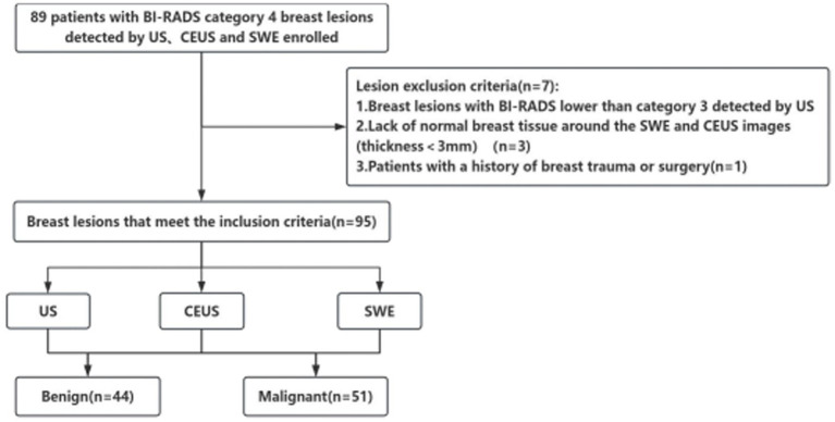
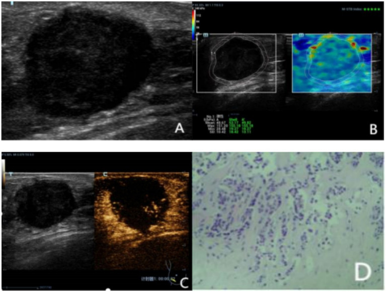
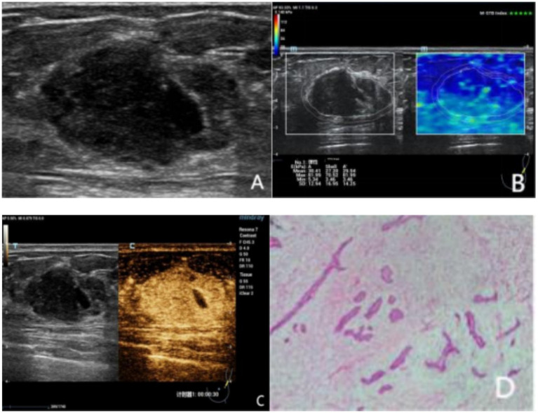
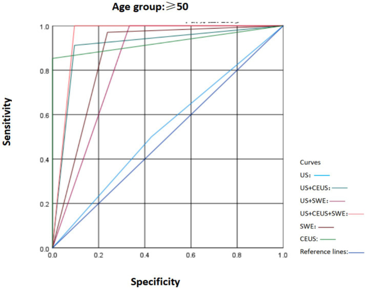
Materials and methods: From February 2022 to November 2024, 95 patients aged 20 to 90 years with breast diseases, all of whom were female, were included. These lesions were diagnosed as BI-RADS 4 breast lesions by conventional ultrasound. All lesions were pathologically confirmed by surgical resection or tissue biopsy, and they were further evaluated by CEUS and SWE. The sensitivity, specificity, positive predictive value (PPV), negative predictive value (NPV), and accuracy of US, CEUS, and SWE were statistically analyzed, and ROC curves were generated. The diagnostic efficacy of US, US + SWE, US + CEUS, and US + CEUS + SWE were subsequently compared, with the pathology results used as the reference standard.
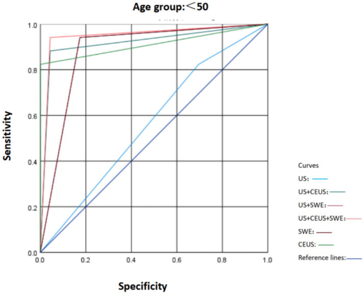
Results: (1) Among the 95 BI-RADS 4 lesions, 44 (46.31%) were benign, and 51 (53.69%) were malignant. The sensitivity, specificity, PPV, NPV and accuracy of the BI-RADS classification via conventional US were 86.3, 72.7, 78.6, 82.1 and 80.0%, respectively. (2) The sensitivity, specificity, PPV, NPV, and accuracy of US combined with SWE in the diagnosis of breast nodules were 96.1, 79.5, 84.5, 94.6, and 88.4%, respectively. (3) The sensitivity, specificity, PPV, NPV, and accuracy of US combined with CEUS in the diagnosis of breast nodules were 84.3, 86.4, 87.8, 82.6, and 85.3%, respectively. (4) The areas under the ROC curve (AUCs) of US, US + SWE, and US + CEUS were 0.795, 0.877, and 0.917, respectively. Statistical methods were used to evaluate the US + CEUS + SWE method, and the results indicated excellent diagnostic performance. The AUC was 0.946, while the sensitivity, specificity, PPV, NPV, and accuracy were 90.7, 93.2, 94.2, 95.3, and 94.7%, respectively. In this this study, the AUCs of US, SWE, and CEUS were compared, and the results revealed that both SWE and CEUS could increase the AUC for breast lesion diagnosis with good diagnostic performance. These methods can increase the sensitivity, specificity and accuracy of the US examination when combined with conventional US. Moreover, the diagnostic performance for breast lesions was highest with the combined application of the three modalities, with a diagnostic AUC that was significantly higher than those of US alone, US + SWE and US + CEUS. The differences were significant (p < 0.05).
Conclusion: (1) CEUS and SWE provide diagnostic information about the microvascular perfusion and tissue stiffness of lesions, respectively, which can assist in the differentiation of benign from malignant breast tumors by conventional US and improve the sensitivity, specificity and accuracy of diagnosis, especially for US BI-RADS 4a breast lesions. (2) The combined use of CEUS and SWE in conventional US enhance the overall diagnostic performance with respect to breast lesions, with the best sensitivity and specificity and the highest diagnostic efficacy. The use of US + CEUS + SWE is beneficial for further differentiating benign and malignant breast lesions according to the US BI-RADS 4, thereby reducing unnecessary biopsies or surgeries.
Keywords: breast tumor; contrast-enhanced ultrasound; multimodal ultrasound examination; shear-wave elastography; ultrasonography.
Copyright © 2025 Li, Wu and Haiqing.
Conflict of interest statement
The authors declare that the research was conducted in the absence of any commercial or financial relationships that could be construed as a potential conflict of interest.
Figures

References
-
- Nicosia L, Bozzini AC, Palma S, Pesapane F, Meneghetti L, Pizzamiglio M, et al. Breast imaging reporting and data system and contrast enhancement mammography: lesion conspicuity likelihood of malignancy and relationship with breast tumor receptor status. Acad Radiol. (2023) 30:2243–51. doi: 10.1016/j.acra.2023.02.008, PMID: - DOI - PubMed
LinkOut - more resources
Full Text Sources