

Subepithelial Lesions of the Ocular Surface: A Review
- PMID: 40552248
- PMCID: PMC12182479
- DOI: 10.1007/s40135-025-00335-8
Subepithelial Lesions of the Ocular Surface: A Review
Abstract
Purpose of review: Subepithelial lesions of the ocular surface represent a diverse group of pathologies which may be difficult to diagnose clinically. Some of these lesions are relatively uncommon, may result in systemic manifestations, or occur secondary to systemic disease. The purpose of this review is to summarize current approaches to the diagnosis and management of six subepithelial conjunctival lesions.
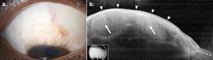
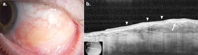
Recent findings: The standard for the diagnosis of subconjunctival lesions remains histopathologic evaluation; however, high-resolution anterior segment optical coherence tomography (HR-OCT) is a useful supplemental diagnostic tool that may facilitate diagnosis. Recent advancements in the management of subconjunctival lesions include targeted systemic therapies in conjunctival melanoma and ultra-low dose radiation radiotherapy in conjunctival lymphoma.
Summary: The development of HR-OCT has provided clinicians with valuable supplemental diagnostic information to guide the diagnosis of subepithelial lesions. Additionally, novel treatment modalities may provide an alternative to traditional surgical interventions in some pathologies.
Keywords: Benign reactive lymphoid hyperplasia; Conjunctival amyloidosis; Conjunctival lymphoma; Conjunctival melanoma; Conjunctival myxoma; Conjunctival neuroma; High-resolution optical coherence tomography.
© The Author(s) 2025.
Conflict of interest statement
Competing InterestsThis work was supported by the Department of Veterans Affairs, Veterans Health Administration, Office of Research and Development, Clinical Sciences R&D (CSRD) I01 CX002015 (Dr. Galor), Biomedical Laboratory R&D (BLRD) Service I01 BX004893 (Dr. Galor), Department of Defense Gulf War Illness Research Program (GWIRP) W81XWH-20-1-0579 (Dr. Galor) and Vision Research Program (VRP) W81XWH-20-1-0820 (Dr. Galor), National Eye Institute R01EY026174 (Dr. Galor) and R61EY032468 (Dr. Galor), NIH Center Core Grant P30EY014801 (institutional), Research to Prevent Blindness Unrestricted Grant GR004596 (institutional), The Dr. Ronald and Alicia Lepke Grant, The Lee and Claire Hager Grant, The Robert Farr Family Grant, The Grant and Diana Stanton-Thornbrough, The Robert Baer Family Grant, The Roberto and Antonia Menendez Grant, The Emilyn Page and Mark Feldberg Grant, The Calvin and Flavia Oak Support Fund, The Jose Ferreira de Melo Grant, The Richard and Kathy Lesser Grant, The Honorable A. Jay Cristol Grant, The Michele and Ted Kaplan Grant, The Carol Soffer Grant, The Richard Azar Family Grant, The Mr. and Mrs. Irwin Friedman Grant, The Dr. Tim and Cammy Ioannides Grant, The Stephen Takach Grant, The Ragheb Family Grant, and The Zvi Levin Grant (institutional grants). Dr. Galor is a Section Editor for Current Ophthalmology Reports. All other authors declare that they have no competing interests.
Figures
References
-
- Simpson T, Fonn D. Optical coherence tomography of the anterior segment. Ocul Surf. 2008;6(3):117–27. 10.1016/s1542-0124(12)70280-x. - PubMed
-
- Shields CL, Yaghy A, Dalvin LA, Vaidya S, Pacheco RR, Perez AL, et al. Conjunctival melanoma: features and outcomes based on the Fitzpatrick skin type in 540 patients at a single ocular oncology center. Ophthalmic Plast Reconstr Surg. 2020;36(5):490–6. 10.1097/IOP.0000000000001624. - PubMed
-
- Shields CL, Shields JA, Gündüz K, Cater J, Mercado GV, Gross N, et al. Conjunctival melanoma: risk factors for recurrence, exenteration, metastasis, and death in 150 consecutive patients. Arch Ophthalmol. 2000;118(11):1497–507. 10.1001/archopht.118.11.1497. - PubMed
Publication types
LinkOut - more resources
Full Text Sources
Research Materials