

How do different cell populations orchestrate myelin regeneration?
- PMID: 40552465
- PMCID: PMC12312399
- DOI: 10.1042/BST20231085
How do different cell populations orchestrate myelin regeneration?
Abstract
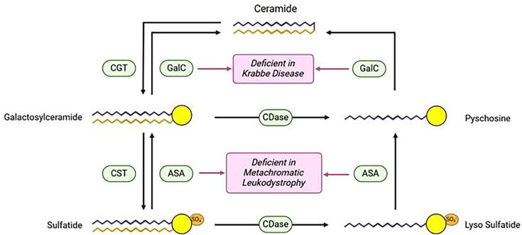
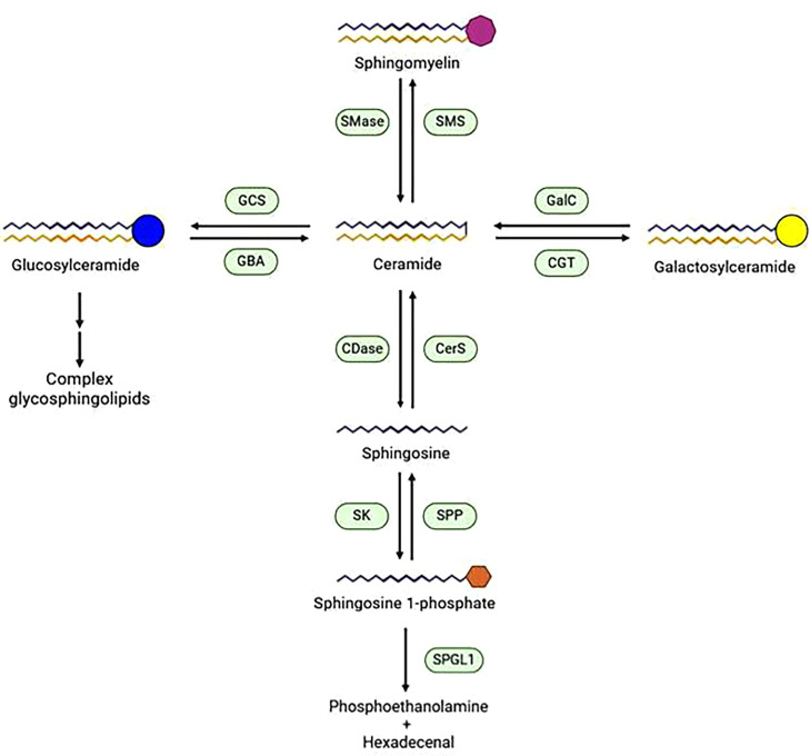
Approximately 35 in 100,000 people are affected by diseases associated with loss of myelin, generally described as demyelinating diseases. Demyelinating diseases encompass many different pathological conditions characterized by heterogeneous and sometimes disease-specific etiopathological mechanisms. While several approaches aimed at ameliorating the symptoms and the progression of some of these diseases exist, the most effective cure for all demyelinating diseases would be regeneration of lost myelin. Myelin regeneration occurs spontaneously in the central nervous system in response to myelin damage but is inefficient for a variety of reasons, especially in human patients. In this review, we will discuss the contributions of different cell populations to the creation of conditions permissive for effective remyelination and to the formation of new myelin after injury. Moreover, we would like to highlight the importance of sphingolipids in the network of interactions between these cell populations. Mutations in genes encoding sphingolipid metabolic enzymes (such as GALC) represent a major risk factor for multiple sclerosis, and alterations in sphingolipid metabolism in specific cell types contribute to myelin damage. On the other hand, sphingolipid signaling, in particular through sphingosine 1 phosphate, directly affects the process of myelin regeneration, with distinct effects on different cellular populations.
Keywords: multiple sclerosis; rHIgM22; remyelination; sphingolipids; sphingosine 1-phosphate.
© 2025 The Author(s).
Conflict of interest statement
The Author declares that there are no competing interests associated with the manuscript.
Figures
Similar articles
-
Uncommon Non-MS Demyelinating Disorders of the Central Nervous System.Curr Neurol Neurosci Rep. 2025 Jul 1;25(1):45. doi: 10.1007/s11910-025-01432-8. Curr Neurol Neurosci Rep. 2025. PMID: 40591029 Review.
-
The Black Book of Psychotropic Dosing and Monitoring.Psychopharmacol Bull. 2024 Jul 8;54(3):8-59. Psychopharmacol Bull. 2024. PMID: 38993656 Free PMC article. Review.
-
A rapid and systematic review of the clinical effectiveness and cost-effectiveness of paclitaxel, docetaxel, gemcitabine and vinorelbine in non-small-cell lung cancer.Health Technol Assess. 2001;5(32):1-195. doi: 10.3310/hta5320. Health Technol Assess. 2001. PMID: 12065068
-
Short-Term Memory Impairment.2024 Jun 8. In: StatPearls [Internet]. Treasure Island (FL): StatPearls Publishing; 2025 Jan–. 2024 Jun 8. In: StatPearls [Internet]. Treasure Island (FL): StatPearls Publishing; 2025 Jan–. PMID: 31424720 Free Books & Documents.
-
Low-Intensity Physical Exercise is Associated with Improved Myelination and Reduced Microglial Activation in a Cuprizone-Induced Demyelination Model.Neurochem Res. 2025 Jun 5;50(3):182. doi: 10.1007/s11064-025-04441-8. Neurochem Res. 2025. PMID: 40471423 Free PMC article.
References
-
- Henríquez K., Molt F., Gajardo J., Cortés B., Ramirez-Santana M Sociodemographic and clinical characteristics of people with multiple sclerosis and neuro-myelitis optica spectrum disorder in a central northern region of Chile: a prevalence study. Mult. Scler. Relat. Disord. 2022;61:103750. doi: 10.1016/j.msard.2022.103750. - DOI - PubMed
Publication types
MeSH terms
Substances
LinkOut - more resources
Full Text Sources