. 2025 Jul 22;99(7):e0007425.
doi: 10.1128/jvi.00074-25. Epub 2025 Jun 24.

CypA inhibits respiratory syncytial virus (RSV) replication by suppressing glycolysis through the downregulation of PKM2 expression

Affiliations

CypA inhibits respiratory syncytial virus (RSV) replication by suppressing glycolysis through the downregulation of PKM2 expression

Jing Zhang et al. J Virol. .

Abstract

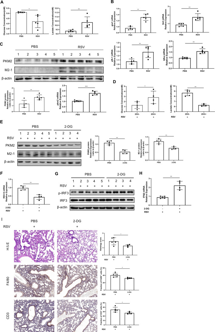
The "Warburg effect," a type of metabolic reprogramming characterized by enhanced glycolysis even in the presence of oxygen, is frequently observed in tumor cells and has also been detected in cells infected with viruses. Our study demonstrated that respiratory syncytial virus (RSV) infection induced aerobic glycolysis both in vivo and in vitro. By utilizing the glycolysis agonist PS48 or inhibitor 2-DG, we ascertained that RSV can utilize glycolysis to promote its replication. Mechanistically, glycolysis may facilitate RSV replication by negatively regulating the IFNβ response. Additionally, we discovered a host molecule, namely CypA, that could downregulate glycolysis to combat RSV infection. CypA interacted with PKM2, a key enzyme of glycolysis, and reduced its expression. By overexpressing or knocking down CypA, we verified that CypA could inhibit aerobic glycolysis, enhance IFNβ production, and reduce RSV replication. Inhibiting the PPIase activity of CypA resulted in the disappearance of its function, indicating that CypA exerted its effects dependent on PPIase activity. Furthermore, we found that CypA has a synergistic effect with 2-DG and an antagonistic effect with PS48 on the IFNβ response, supporting the notion that CypA regulates IFNβ by inhibiting glycolysis. These results indicate that CypA may serve as a novel host factor in the regulation of glycolysis, the interferon response, and ultimately in resisting RSV infection.

Importance: Viruses utilize the host's resources and energy to carry out essential life processes and achieve self-replication. In response, hosts have evolved a range of antagonistic mechanisms. Our study investigates how RSV employs glycolysis to benefit its replication, with a particular focus on the interaction between glycolysis and IFNβ regulation. Additionally, we explore how the host employs CypA to antagonize the virus's utilization of glycolysis, thereby inhibiting RSV replication. Our findings will contribute to the development of effective antiviral therapies targeting CypA.

Keywords: CypA; IFNβ; PKM2; RSV; glycolysis; viral replication.


Conflict of interest statement

The authors declare no conflict of interest.

Figures

Fig 1
Fig 1
RSV infection induced aerobic glycolysis. (A, F, and G) HEp-2 cells were infected with RSV-GFP (MOI = 1) for indicated times. (A) Glucose concentration and lactate concentration of cell culture supernatant in the mock group and infection group at different times were detected by relevant assay kits. (B–E) HEp-2 cells were infected with RSV-GFP (MOI = 1) for 24 h. GLUT1, HK2, PFK-1, and LDHA gene expressions were detected by qRT-PCR. (F and G) PKM2 expression at different times of infection was detected by qRT-PCR or WB. (H and I) Anticoagulant blood samples from the clinic. (H) Plasma glucose and lactate concentrations were detected by relevant assay kits. (I) PKM2 gene expression in PBMCs was detected by qRT-PCR. Data represent the mean ± SD of three independent experiments. (n = 3, *P < 0.05, **P < 0.01, ***P < 0.001, ****P < 0.0001).
Fig 2
Fig 2
Glycolysis promotes RSV replication by limiting interferon production. HEp-2 cells were infected with RSV-GFP (MOI = 1) in the absence or presence of 1 mM 2-DG (A–D, K and L) or 20 µM PS48 (A and B, E and F, K and L). (G–J, M and N) After transfecting with si-Ctrol or si-LDHA for 48 h, HEp-2 cells were infected with RSV-GFP (MOI = 1) for 24 h. (A, G) The concentrations of glucose and lactate in the supernatant of cell culture were determined by relevant assay kits for indicated times. (B, H) Expressions of PKM2 and M2-1 were determined by WB. (C, E, I) Fluorescence images of RSV-GFP replication in HEp-2 cells treated with 2-DG (C) or PS48 (E) were transfected with si-LDHA (I), and the mean fluorescence intensity was quantified, the uninfected group served as the control group. (D, F, J) RSV-N mRNA expression was detected by qRT-PCR. Viral titers were detected by viral plaque assay. (K and L) HEp-2 cells treated with or without 2-DG or PS48 were stimulated with Poly (I:C). (M and N) After transfecting with si-Ctrol or si-LDHA for 48 h, cells were stimulated with Poly (I:C). (K, M) IRF3 phosphorylation levels were detected by WB. (L, N) IFNβ mRNA expression was determined by qRT-PCR. Data represent the mean ± SD of three independent experiments (n = 3, *P < 0.05, **P < 0.01, ***P < 0.001).
Fig 3
Fig 3
Glycolysis is essential for the replication of RSV in vivo. (A–C) C57/B6 mice were infected with RSV (5 × 10⁶ PFU per mouse) for 3 days. (A) Mice serum was collected for the detection of glucose and lactate concentrations. (B) GLUT1, HK2, PFK-1, and LDHA gene expressions in mouse lungs were detected by qRT-PCR. (C) PKM2 expression in mouse lungs was detected by WB or qRT-PCR. (D–G) C57/B6 mice were infected with RSV (5 × 10⁶ PFU per mouse) for 3 days. Mice were intraperitoneally injected with 2-DG (1,000 mg/kg) or PBS for three consecutive days from the first day of infection. (D) Mice serum was collected for the detection of glucose and lactate concentrations. (E) Expressions of PKM2 and M2-1 in mouse lungs were determined by WB. (F) RSV-N mRNA expression in mouse lungs was detected by qRT-PCR. IRF3 phosphorylation level in mouse lungs were detected by WB (G), and IFNβ mRNA expression was determined by qRT-PCR (H). (I) Pathological examination of mouse lungs: HE staining and immunohistochemical detection of F4/80 and CD3. Data represent the mean ± SD of three independent experiments (n = 3, *P < 0.05, **P < 0.01, ***P < 0.001). Results in vivo are means ± SD for five mice per group.
Fig 4
Fig 4
CypA downregulates PKM2 expression to inhibit aerobic glycolysis during RSV infection. (A) 24 h after transfecting indicated plasmids, HEK293T cells were infected with RSV-GFP for 24 h. Co-IP was performed, and then, WB was done to identify the interaction between CypA and PKM2. (B–E) 24 h after transfecting with Myc-vector or Myc-CypA plasmid, HEp-2 cells were infected with RSV-GFP for 24 h. (F–I) After transfecting with si-Ctrol or si-CypA for 48 h, HEp-2 cells were infected with RSV-GFP for 24 h. (B, F) The replication of RSV-GFP was observed by fluorescence microscope, and the mean fluorescence intensity was quantified; the uninfected group served as the control group. (C, G) Viral titers were detected by viral plaque assay. (D, H) PKM2 mRNA and protein expressions were detected by qRT-PCR and WB, M2-1 protein expression was detected by WB. (E–I) The concentrations of glucose and lactate in the supernatant of cell culture were determined by relevant assay kits. (J) HEp-2 cells were transfected with the indicated plasmids. 24 h post-transfection, cells were infected with RSV for 24 h in the presence of bafilomycin A1 or treated with MG132 for 12 h before collection. Data represent the mean ± SD of three independent experiments (n = 3, *P < 0.05, **P < 0.01, ***P < 0.001).
Fig 5
Fig 5
CypA could positively regulate IFNβ response, which have a synergistic effect with 2-DG and an antagonistic effect with PS48. 24 h after transfecting with Myc-vector or Myc-CypA plasmid (A), 48 h after transfecting with si-Ctrol or si-CypA (C), HEp-2 cells were infected with RSV-GFP for 24 h. Phosphorylation of IRF3 or IFNβ mRNA expression was detected by WB or qRT-PCR. (B, D) After transfection with Myc-CypA (B) or si-CypA (D), HEp-2 cells were transfected with 1 µg/mL Poly(I:C). The concentration of IFNβ in cell culture supernatant was determined by ELISA. (E, G) HEp-2 cells were transfected with indicated plasmids for 24 h and then transfected with 1 µg/mL Poly(I:C) for 24 h. Add 2-DG (E) or PS48 (G) when changing the medium. (F, H) HEp-2 cells were transfected with indicated siRNA for 48 h and then transfected with 1 µg/mL Poly(I:C) for 24 h. Add 2-DG (F) or PS48 (H) when changing the medium. (E–H) Phosphorylation of IRF3 or IFNβ mRNA expression was detected by WB or qRT-PCR. Data represent the mean ± SD of three independent experiments (n = 3, *P < 0.05, **P < 0.01, ***P < 0.001).
Fig 6
Fig 6
CypA inhibits glycolysis and promotes interferon production in HEp-2 cells dependent on its peptidyl-prolyl cis-trans isomerase activity. (A–E) HEp-2 cells were transfected with Myc-vector, Myc-CypA, or Myc-CypA-R55A for 24 h and then infected with RSV for 24 h. (A) The replication of GFP-RSV was observed by fluorescence microscope, and the mean fluorescence intensity was quantified; the uninfected group served as the control group. (B) Viral titers were detected by viral plaque assay. (C) PKM2 mRNA and protein expressions, M2-1 protein expression was detected by qRT-PCR or WB. (D) The concentrations of glucose and lactate in the supernatant of cell culture were determined by relevant assay kits. (E) Phosphorylation of IRF3 and IFNβ gene expression were detected by WB or qRT-PCR. (F) After transfection with indicated plasmids for 24 h, HEp-2 cells were transfected with 1 µg/mL Poly(I:C). The concentration of IFNβ in cell culture supernatant was determined by ELISA kit. (G–L) 4 h after infecting with RSV, HEp-2 cells were treated with DMSO or CsA for 24 h. (G) The replication of GFP-RSV was observed by fluorescence microscope, and the mean fluorescence intensity was quantified, uninfected group served as the control group. (H) Viral titers were detected by viral plaque assay. (I) PKM2 mRNA and protein expressions, M2-1 protein expression was detected by qRT-PCR or WB. (J) The concentrations of glucose and lactate in the supernatant of cell culture were determined by relevant assay kits. (K) Phosphorylation of IRF3 and IFNβ gene expression were detected by WB or qRT-PCR. (L) After transfecting with 1 µg/mL Poly(I:C), HEp-2 cells were treated with DMSO or CsA for 24 h. The concentration of IFNβ in cell culture supernatant was determined by ELISA kit. Data represent the mean ± SD of three independent experiments (n = 3, ns P > 0.05, *P < 0.05, **P < 0.01, ***P < 0.001).

Similar articles

References

    1. Blanco JCG, Boukhvalova MS, Hemming P, Ottolini MG, Prince GA. 2005. Prospects of antiviral and anti-inflammatory therapy for respiratory syncytial virus infection. Expert Rev Anti Infect Ther 3:945–955. doi: 10.1586/14787210.3.6.945 - DOI - PubMed
    1. Hoffman SJ, Laham FR, Polack FP. 2004. Mechanisms of illness during respiratory syncytial virus infection: the lungs, the virus and the immune response. Microbes Infect 6:767–772. doi: 10.1016/j.micinf.2004.03.010 - DOI - PubMed
    1. Battles MB, McLellan JS. 2019. Respiratory syncytial virus entry and how to block it. Nat Rev Microbiol 17:233–245. doi: 10.1038/s41579-019-0149-x - DOI - PMC - PubMed
    1. Coultas JA, Smyth R, Openshaw PJ. 2019. Respiratory syncytial virus (RSV): a scourge from infancy to old age. Thorax 74:986–993. doi: 10.1136/thoraxjnl-2018-212212 - DOI - PubMed
    1. Wright M, Piedimonte G. 2011. Respiratory syncytial virus prevention and therapy: past, present, and future. Pediatr Pulmonol 46:324–347. doi: 10.1002/ppul.21377 - DOI - PubMed

MeSH terms

LinkOut - more resources