

The Influence of Deep Margin Elevation and Immediate Dentin Sealing on the Fracture Strength of Premolars Restored With Indirect Inlays: An In Vitro Study
- PMID: 40553010
- PMCID: PMC12186461
- DOI: 10.1002/cre2.70161
The Influence of Deep Margin Elevation and Immediate Dentin Sealing on the Fracture Strength of Premolars Restored With Indirect Inlays: An In Vitro Study
Abstract
Objective: To evaluate the effect of deep margin elevation (DME) and immediate dentin sealing (IDS) on the fracture strength of premolars restored with lithium disilicate inlay restorations.
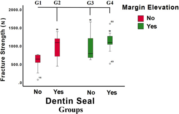
Materials and methods: Standard MOD inlays with proximal box preparations extending 3 mm apical to the cementoenamel junction were prepared on forty sound premolars (n = 10) randomly divided into four groups: inlays without DME and without IDS (G1); inlays without DME but with IDS (G2); inlays with DME but without IDS (G3); and inlays with both techniques applied (G4). Composite resin was applied incrementally to elevate the proximal cervical margin coronally to the cementoenamel junction. For immediate dentin sealing, bonding agent was applied immediately after tooth preparation. All teeth were restored with lithium disilicate inlays and, after adhesive resin cementation, specimens were thermomechanically aged for 500 cycles at 5°-55°C and then subjected to load failure testing. Failure loads and locations were recorded and analyzed using one- and two-way ANOVA with Tukey's post-hoc testing (α = 0.05). Failure modes were analyzed using descriptive statistics.
Results: The mean fracture loads were 565.76 ± 233.22 N, 978.47 ± 394.2 N, 974.31 ± 334.7 N, and 1108.21 ± 292.41 N for G1, G2, G3, and G4, respectively. Deep margin elevation (p = 0.011) and immediate dentin sealing (p = 0.010) were associated with significantly increased fracture loads. Fracture loads were significantly lower for G1 teeth than for G2-G4 teeth, but there were no significant differences between G2, G3, and G4. G1 teeth showed 50% catastrophic and non-catastrophic failures, which increased to 60% for G2 and decreased to 20% for G3 teeth. Samples with both seals and elevation (G4) had a 60% catastrophic failure rate.
Conclusions: When applied individually or together, deep margin elevation and immediate dentin sealing significantly increase the fracture strength of premolars restored with indirect lithium disilicate inlays.
Clinical implications: In the challenging setting of margin elevation, studies on the effects of immediate dentin sealing have generally been limited to evaluating marginal integrity and bond strength. The findings of this In Vitro study suggest that both deep margin elevation and immediate dentin sealing protocols are likely to improve clinical outcomes of indirect lithium disilicate inlay restorations and may be considered viable options in clinical practice.
Keywords: deep margin elevation; dentin sealing; fracture strength; lithium disilicate inlay restorations.
© 2025 The Author(s). Clinical and Experimental Dental Research published by John Wiley & Sons Ltd.
Conflict of interest statement
The authors declare that they have no known competing financial interests or personal relathioships that could have appeared to influence the work reported in this paper.
Figures


References
-
- Bertschinger, C. , Paul S. J., Lüthy H., and Schärer P.. 1996. “Dual Application of Dentin Bonding Agents: Effect on Bond Strength.” American Journal of Dentistry 9, no. 3: 115–119. - PubMed
MeSH terms
Substances
Grants and funding
LinkOut - more resources
Full Text Sources
