. 2025 Jun 24;10(15):e192929.
doi: 10.1172/jci.insight.192929. eCollection 2025 Aug 8.

LRP2 is a potential molecular target for nonsyndromic pathological myopia

Affiliations

LRP2 is a potential molecular target for nonsyndromic pathological myopia

Kimberley Delaunay et al. JCI Insight. .

Abstract

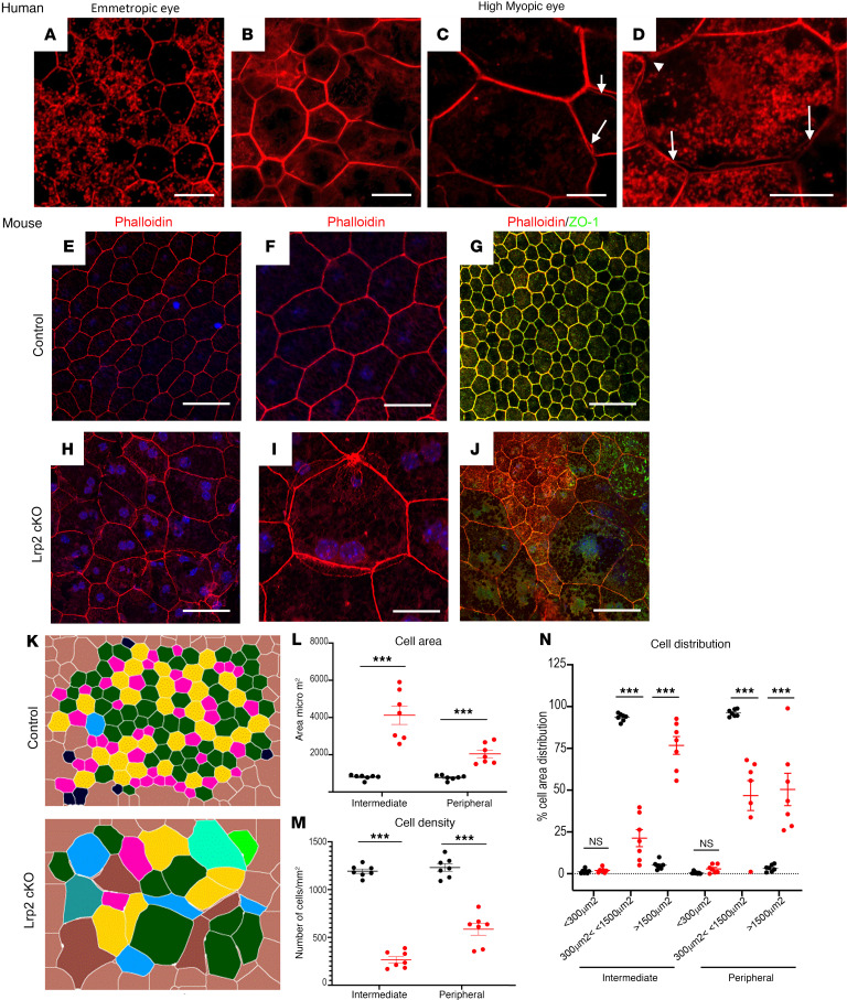
High myopia (HM) and posterior staphyloma (PS) are major causes of vision loss worldwide. Genetic and environmental factors, especially light exposure, influence myopia. This study shows that low-density lipoprotein-related receptor type 2 (LRP2) levels are decreased in the vitreous of patients with HM and PS, and that in human donor eyes affected by PS, LRP2 expression was reduced in the neural retina and retinal pigment epithelium (RPE), with morphologic changes similar to those observed in the Foxg1-Cre-Lrp2fl/fl mouse that also develops PS. In human induced pluripotent stem cell-derived RPE cells, LRP2 silencing regulated genes involved in eye and neuronal development, visual perception, tissue remodeling, hormone metabolism, and RPE structure. Its expression increased under light exposure, particularly red light, but was downregulated by cortisol. These findings establish a link between LRP2, myopization, and environmental factors, highlighting its crucial role in nonsyndromic HM and PS. LRP2 appears to be a promising therapeutic target for HM treatment.

Keywords: Neuroscience; Ophthalmology; Retinopathy.


Figures

Figure 1
Figure 1. Analysis of proteins in the vitreous of myopic and emmetropic patients.
(A) Volcano plot of differentially expressed proteins. The rightmost part of the plot (blue circles) shows the 20 upregulated proteins and the leftmost (red circles) the 53 downregulated proteins. PM, pathological myopia. (B) Monarch analysis (https://monarchinitiative.org/) enrichment of downregulated proteins; only the top 5 terms are displayed. Gene count associated with a particular Monarch term is indicated. (C) Protein-protein interactions (PPIs) using STRING analysis (see Supplemental Methods), displaying the signaling network between the differentially expressed proteins. Nodes in blue circles refer to upregulated proteins, nodes in gray circles refer to downregulated proteins. Markov cluster algorithm analysis indicated the main interactomes. The 9 top interactomes are indicated at the right. (D) Concentrations of LRP2 protein in ng/mL in undiluted vitreous from control (n = 13) and NSHM (n = 10) eyes quantified with ELISA. The graph on the right shows the ratio of LRP2 compared to the total protein content. Statistical analysis was performed using the Mann-Whitney U test. *P < 0.05.
Figure 2
Figure 2. Ophthalmologic imaging and histological analysis of the right eye of an HM donor.
(A and B) Premortem SD-OCT scans of the right eye with inferior cross section (A) and macular cross section (B), showing the posterior staphyloma (PS) (white arrows) and tractional maculopathy (yellow stars). The myopic eye displayed an atrophy of the macula delineated by small arrows and the posterior incurvation indicated the uncomplicated PS. (C) Gross morphology of the HM right enucleated postmortem eye with PS. (D) Macroscopic image of the posterior chamber after dissection showing the PS. (E) Insert box of D focusing on fovea and PS (black arrow). (F) DAPI-stained section of a normal human retina near the macula. (G) DAPI-stained section of the HM retina with a PS (double headed arrow) shows a severe reduction in thickness of all retinal layers, adjacent to the PS. Schisis (white arrow) is observed between the inner nuclear layer (inl) and the outer nuclear layer (onl). A cyst is observed between the RPE and the outer segments layer (asterisk). bv, blood vessel. (H) RPE cells in human retina form a single layer of pigmented cells as observed on light transmission microscopy. Melanosomes (m) have an elongated shape and nuclei (n) are spaced at regular intervals. (I and J) In the HM donor eye, RPE cells are still forming a single layer. RPE cells are abnormally large, their apical domain is reduced, and melanosomes aggregate in macromelanosomes (mm) or macrostructures (ag). Scale bars: 75 μm (F), 120 μm (G), and 5 μm (HJ).
Figure 3
Figure 3. Immunolabeling of retinal cells in the emmetropic and HM retina.
(A and B) Glutamine synthetase (GS) labels Müller glial cells in the emmetropic retina, extending radially from the nerve fiber layer (nfl) to the outer nuclear layer (onl). (C and D) In HM retina, GS-positive cells extend in all directions and thicken at the retinal surface. At higher magnification, GS-positive cells surround a cyst in the ganglion cell layer (gcl) (C) and fill the bottom of the staphyloma where almost no retina remains (E). (F and G) Tubulin β3 (Tubb3) is a neuronal marker of retinal ganglion (RGC) and amacrine cells in the emmetropic retina. (H) In HM retina, the number of Tubb3-positive cells and RGCs is reduced. Higher magnification shows Tubb3-positive axons browsing at the surface of the staphyloma. (J and K) Blue (red staining) and green (green staining) opsin markers reveal the distribution of blue and green cones in the emmetropic retina. (L and M) In HM retina, the number of blue cones is reduced (L) and high magnification shows the accumulation of green opsin in abnormally shaped outer segments. (N and O) Glial fibrillary acidic protein (GFAP) labels astrocytes and glial Müller cell endfeet in the emmetropic retina. (P and Q) In the HM retina, the GFAP-positive endfeet are reduced. (Q) At higher magnification, a GFAP-positive membrane lays on the retinal surface but no positive cells are observed in the retina. Scale bars: 200 μm (A, F, J, and N), 80 μm (B, G, and O), 20 μm (K), 250 μm (C, H, L, and P), and 60 μm (D, E, I, M, and Q).
Figure 4
Figure 4. LRP2 immunolabeling in the emmetropic and HM retina.
(A and B) In the emmetropic neural retina, LRP2 is expressed around RGCs and along GS-positive glial Müller cells. (C and D) In the HM neural retina, LRP2 expression is greatly reduced in GS-positive cells, which surround cysts (stars). (E) In transversal sections of RPE cells, LRP2 is located at the apical pole (white arrowheads), in intracellular vesicles, and at the basal pole of the RPE, and in the pillars of the choriocapillaris. (F and G) In an RPE flat-mounted preparation from the left emmetropic eye, LRP2 is distributed in cytoplasmic vesicles (F) along apical and lateral membranes (G) and most LRP2-positive vesicles are also positive for clathrin. (I) In transversal section of the HM RPE, LRP2 expression is greatly diminished and absent in the choriocapillaris. (JL) In RPE flat mounted from the HM eye, LRP2 distribution is sparse and diffuse, and does not colocalize with clathrin (L) that is also diminished. Scale bars: 200 μm (AD), 10 μm (E and I), and 50 μm (F, G, and JL).
Figure 5
Figure 5. Organization of the RPE is altered in HM eye, similar to the RPE in Lrp2-cKO mice.
(A) Phalloidin staining reveals the geometric paving pattern of RPE in the emmetropic eye. (BD) In HM eye, RPE geometric paving pattern is lost as most cells increase in size. Bicellular junctions display secondary actin arcs (C, arrows) and cell junctions are disorganized (D arrows). (E and F) Phalloidin staining on RPE flat mount of WT control mouse shows the regular pattern of RPE cells. (G) Zonula occludens 1 (ZO-1) follows the distribution of phalloidin in the apical pole. (H and I) In Lrp2-cKO RPE, the pseudogeometric paving pattern is lost and is replaced by a tangle of cells whose surface has increased. (J) ZO-1 is redistributed in the cytoplasm of abnormally shaped RPE cells. (K) Digital overlay reconstruction of control and Lrp2-cKO RPE indicates the increase size in cells both at the periphery and at the intermediate level of the retina in Lrp2-cKO RPE. (L) Cell area in μm2, (M) cell density in number of cells/mm2, and (N) distribution of cells by sizes in the intermediate and peripheral retina of control compared to Lrp2-cKO mice. Values represent the mean of cell size average of each sample (n = 7) per genotype ± SEM. Mann-Whitney U test. ***P < 0.001. Scale bars: 50 μm (AD, E, and H), 25 μm (F and I), and 75 μm (G and J).
Figure 6
Figure 6. LRP2 localization in healthy human iRPE.
(A) Consecutive confocal images from an apical to basal stack showing LRP2 accumulation in large vesicular structures in the apical pole (arrows), at lateral membranes, in small cytoplasmic vesicles, and in larger perinuclear vesicles (arrowheads). (B) Consecutive confocal images from an apical to basal stack show that LRP2 colocalized with the major protein of coated pits and apical endocytic vesicles, clathrin (yellow arrow). In the basal part of the cell, LRP2 (green arrow) did not colocalize with clathrin (red arrow). (C) Confocal image showing that LRP2 (green) partially colocalized with the endocytic marker, early endosome antigen 1 (EEA1; red) (yellow arrow). (D) In the basal compartment, LRP2 (green) colocalized with LAMP1 (red), a specific lysosomal marker (yellow arrows). Scale bars: 10 μm (A, C, and D) and 14 μm (B).
Figure 7
Figure 7. Differentially expressed genes (DEGs) between siLRP2 and scrambled iRPE cells.
(A) Lrp2 mRNA quantification between scrambled (n = 4) and siLRP2 (n = 4) iRPE (*P < 0.05). (B) LRP2 levels as expressed by the ratio LRP2/GAPDH between scrambled (n = 4) and siLRP2 (n = 4) iRPE (**P < 0.01). (C) Volcano plot of DEGs between siLRP2 and scrambled iRPE cells with –log10 of the adjusted P value on the y axis and log2 of the fold change in expression on the x axis. Some genes, including LRP2, are indicated. (D) Category netplot gene enrichment analysis considering only LRP2 as a common factor with 3 GO terms (sensory perception, import across plasma membrane, and steroid metabolic processes) and (E) with 2 Reactome terms (sensory perception, visual transduction and metabolism). Fold change (color codes on each graph) for each gene of the selected GO term is indicated. Analysis was performed using Metascape (https://metascape.org/). Absolute normalized enrichment allowed identifying terms that were upregulated or downregulated using all DEGs.
Figure 8
Figure 8. LRP2 expression and environmental factors.
(A) Quantification of LRP2 mRNA by qPCR in iRPE not exposed (ne) or exposed to light (left panel) after 0.5, 2, or 10 hours after illumination. Left panel showing quantification of LRP2 mRNA in iRPE not exposed or exposed to light. Right panel showing quantification of LRP2 mRNA in iRPE not exposed or exposed to red, blue, or white light. Values correspond to the means of 4 independent experiments in duplicate for each condition. Each independent experiment represents the mean of 3 wells. Data are expressed as fold change in gene expression ± SD and were analyzed using the nonparametric Kruskal-Wallis test and Mann-Whitney post hoc test. *P < 0.05; **P < 0.01; #P > 0.05 (not significant). (B) LRP2 and EEA1 expression in iRPE not exposed or exposed to red light. (C) LRP2 mRNA expression in iRPE cultures treated without (ethanol) or cortisol. Values correspond to the mean of 6 experiments in triplicate. Data represent the mean fold change in gene expression ± SEM. Mann-Whitney U test. *P < 0.05. Scale bars: 20 μm (LRP2) and 40 μm (EAA1).

Similar articles

References

    1. Wolffsohn JS, et al. IMI-Global trends in myopia management attitudes and strategies in clinical practice-2022 update. Invest Ophthalmol Vis Sci. 2023;64(6):6. doi: 10.1167/iovs.64.6.6. - DOI - PMC - PubMed
    1. Ohno-Matsui K, Jonas JB. Posterior staphyloma in pathologic myopia. Prog Retin Eye Res. 2019;70:99–109. doi: 10.1016/j.preteyeres.2018.12.001. - DOI - PubMed
    1. Ruiz-Medrano J, et al. Myopic maculopathy: current status and proposal for a new classification and grading system (ATN) Prog Retin Eye Res. 2019;69:80–115. doi: 10.1016/j.preteyeres.2018.10.005. - DOI - PubMed
    1. Ruiz-Medrano J, et al. The importance of the type of posterior staphyloma in the development of myopic maculopathy. Diagnostics (Basel) 2024;14(15):1581. doi: 10.3390/diagnostics14151581. - DOI - PMC - PubMed
    1. Shi H, et al. Global prevalence of myopic macular degeneration in general population and patients with high myopia: a systematic review and meta-analysis. Eur J Ophthalmol. 2024;34(3):631–640. doi: 10.1177/11206721231185816. - DOI - PubMed

MeSH terms

Substances

LinkOut - more resources