. 2025 Jul:117:105810.
doi: 10.1016/j.ebiom.2025.105810. Epub 2025 Jun 18.

Identification of miR-106b-5p as a senolytic miRNA

Affiliations

Identification of miR-106b-5p as a senolytic miRNA

Tianpeng Zhang et al. EBioMedicine. 2025 Jul.

Abstract

Background: Cellular senescence contributes to ageing and age-related diseases. While miR-106b-5p is elevated in centenarians and GH-deficient models of healthy ageing, its role in senescence was unclear.

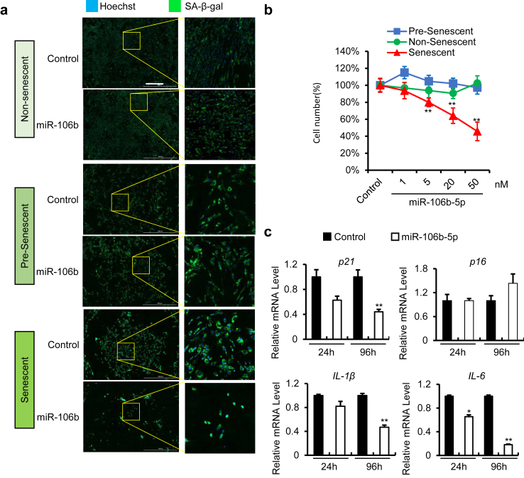
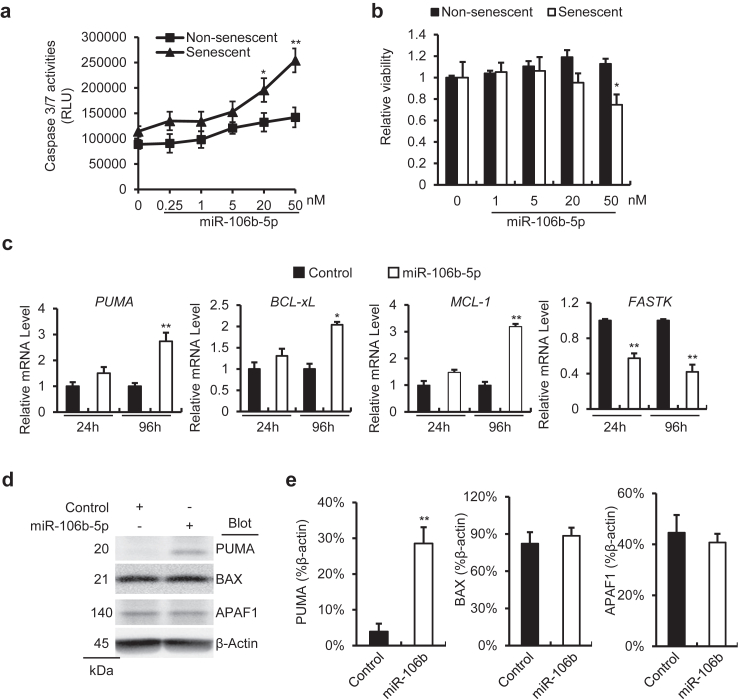
Methods: Senolytic effects of miR-106b-5p were evaluated in etoposide-induced senescent IMR90 fibroblasts and HUVECs, and in male naturally aged mice using liposome-mediated delivery. Cellular assays, qPCR, Western blotting, and RNA-seq were performed to assess senescence and SASP markers, apoptosis pathways, and molecular mechanisms.

Findings: miR-106b-5p selectively eliminated senescent cells without affecting non-senescent cells. It enhanced p53 K120 acetylation and upregulated PUMA, while reducing PCAF expression. In male aged mice, systemic delivery of miR-106b-5p reduced markers of senescence and SASP in multiple tissues and lowered serum IL-6 levels.

Interpretation: miR-106b-5p functions as a senolytic miRNA via modulation of the p53-PUMA axis and SASP suppression. It holds promise as a therapeutic agent to mitigate age-related cellular dysfunction and inflammation.

Funding: Supported by NIH (U19 AG056278, R01 AG063543, P01 AG062413, U54 AG079754, U54 AG076041, R01 AG069819, P01 AI172501), the Glenn Foundation, and NSF grant #2317758.

Keywords: Ageing and inflammation; Cellular senescence; SASP; Senolytic therapy; miR-106b-5p; p53-PUMA axis.


Conflict of interest statement

Declaration of interests Tianpeng Zhang, Allancer Nunes, Ryan O'Kelly, Luise Angelini, Michal M. Masternak, and Yousin Suh declare no competing interests related to this study. Laura J. Niedernhofer holds equity in Unity Biotechnology and has pending IP through the University of Minnesota related to senescence. Paul D. Robbins holds equity in Genascence Inc. and Innate Biologics, stock options in L&J Bio, and has pending patents on senotherapeutics via the University of Minnesota. Xiao Dong is a co-founder and shareholder of SingulOmics Corp. None of these disclosures are related to the present work, and no authors intend to file a patent based on this study.

Figures

Fig. 1
Fig. 1
Selective clearance of senescent IMR90 cells by miR-106b-5p mimic transfection. (a) SA-β-gal senescence analysis using C12FDG of non-senescent, senescent, and pre-senescent IMR90 cells transfected with 50 nM of the miR-106b-5p mimic for 96 h. C12FDG SA-β-gal staining (green) and Hoechst staining (blue), scale bar: 1000 μm (n = 4 per group). Statistical analysis: one-way ANOVA. (b) Quantitative analysis of total cell counts and percentage of senescent cells with increasing doses of the miR-106b- 5p mimic after 96 h treatment (n = 4 per group). Statistical analysis: Student's t-test. (c) qPCR analysis of p21Cip1, p16Ink4, IL-1β, and IL-6 in senescent IMR90 post-transfected with 50 nM miR-106b-5p mimic for 24 and 96 h (n = 4 per group). The senescent control image is reused in both Fig. 1a and Fig. S1b, as the same sample was analysed in parallel experiments. Statistical analysis: Student's t-test. All data represent the mean ± SEM. ∗p < 0.05 and ∗∗p < 0.01 indicate statistical significance.
Fig. 2
Fig. 2
miR-106b-5p selectively eliminates senescent IMR90 cells by inducing senescent cell apoptosis. (a) Caspase 3/7 assay of non-senescent and senescent IMR 90 cells transfected with increasing concentrations of miR-106b-5p mimic for 96 h (n = 3 per group). Statistical analysis: two-way ANOVA. (b) Quantitation of cell viability of non-senescent and senescent IMR90 cells with increasing doses of miR-106b-5p mimic for 96 h using an ATPase assay. Statistical analysis: two-way ANOVA. (c) Expression levels of apoptosis-related genes PUMA, BCL-XL, MCL-1, and FASTK relative to GAPDH quantified by qPCR at 24 and 96 h post-transfection with miR-106b-5p mimic (50 nM, n = 4 per group). Statistical analysis: Student's t-test. (d) Representative immunoblots of senescent IMR90 96 h post-transfection with miR-106b-5p mimic (50 nM). (e) Densitometric quantification of three independent Western blots was performed using ImageJ software. PUMA, BAX, and APAF1 protein expression levels were normalised to β-actin levels in the corresponding samples (n = 3 per group). Statistical analysis: Student's t-test. Data are presented as mean ± SEM. ∗p < 0.05 and ∗∗p < 0.01 denotes statistical significance.
Fig. 3
Fig. 3
miR-106b-5p eliminated senescent cells by upregulating PUMA through K120 acetylation of p53. (a) Analysis of senescence using C12FDG staining for SA-β-gal conducted on senescent IMR90 cells transfected with 25 nM miR-106b-5p mimic with either 25 nM PUMA or a control siRNA, for 96 h (n = 3 per group). Statistical analysis: Student's t-test. (b, c) qPCR analyses of PUMA, p21Cip1, IL-1β, and IL-6 in cells as described in (a). Statistical analysis: Student's t-test. (d) Representative immunoblots of p53 K120, PUMA, BAX, p53 ser15, and p53 at 12 h or 48 h post-transfection with 50 nM miR-106b-5p mimic (n = 3 per group). (e) Densitometry quantification of Western blot bands (n = 3 per group). All treatment groups were compared to the control group in the statistical analyses. Statistical analysis: Student's t-test. Data are shown as mean ± SEM. ∗p < 0.05 and ∗∗p < 0.01 indicate levels of statistical significance.
Fig. 4
Fig. 4
Analysis of the mechanism of the senolytic activity of miR-106b-5p. (a) Gene Set Enrichment Analysis (GSEA) of RNA-seq data was utilised to identify pathways significantly enriched upon miR-106b-5p treatment (n = 3 per group). (b) qPCR analyses of BNIP2, SQSTM1 and PCAF at 24- and 96-h post-transfection with miR-106b-5p mimic (50 nM, n = 4 per group). Statistical analysis: Student's t-test. (c) Assessment of IL-6 levels in the supernatant of senescent IMR90 cells 96-h post-transfection with miR-106b-5p mimic (50 nM). Statistical analysis: Student's t-test. Data represent mean ± SEM. ∗p < 0.05 and ∗∗p < 0.01 denoting statistical significance.
Fig. 5
Fig. 5
Targeting senescence: miR-106b-5p′s role in enhancing health in naturally ageing mice. (a) Representative SA-β-gal staining images from the livers of aged mice treated with three intraperitoneal (I.P.) injections of miR-106b-5p mimics at 2-day intervals; scale bar: 1000 μm (n = 6 per group). (b) Quantitative analysis of SA-β-Gal-positive cells in fresh frozen liver tissue sections (n = 6 per group). (c) Representative immunofluorescence images of γ-H2AX staining (red) with DAPI staining (blue); scale bar: 100 μm (n = 6 per group). (d) Quantification of γ-H2AX staining using Cytation 1 imaging reader. (e) ELISA-based quantification of serum IL-6 levels, indicative of systemic inflammation. (f) qPCR analyses of p16Ink4, P21CIP1, IL-1 β, and IL-6 in the liver of nature ageing mouse. (g) Measurement of endogenous mmu-miR-106b-5p expression levels in the liver of young vs old mice (n = 6 per group). (h) Representative immunoblots of PUMA, p53 k120, p53 in the liver of naturally ageing mice, and (i) densitometric quantification of Western blots were performed using ImageJ software. PUMA, p53 k120, and p53 protein expression levels were normalised to β-actin levels in the corresponding samples (n = 4). (j) Gene Set Enrichment Analysis (GSEA) of RNA-seq data from liver and spleen tissues of aged mice treated with miR-106b-5p. Dot size represents the number of genes in each pathway, and colour indicates the nominal p-value (Nom p-value) of pathway enrichment. Data are presented as mean ± SEM.
Fig. 5
Fig. 5
Targeting senescence: miR-106b-5p′s role in enhancing health in naturally ageing mice. (a) Representative SA-β-gal staining images from the livers of aged mice treated with three intraperitoneal (I.P.) injections of miR-106b-5p mimics at 2-day intervals; scale bar: 1000 μm (n = 6 per group). (b) Quantitative analysis of SA-β-Gal-positive cells in fresh frozen liver tissue sections (n = 6 per group). (c) Representative immunofluorescence images of γ-H2AX staining (red) with DAPI staining (blue); scale bar: 100 μm (n = 6 per group). (d) Quantification of γ-H2AX staining using Cytation 1 imaging reader. (e) ELISA-based quantification of serum IL-6 levels, indicative of systemic inflammation. (f) qPCR analyses of p16Ink4, P21CIP1, IL-1 β, and IL-6 in the liver of nature ageing mouse. (g) Measurement of endogenous mmu-miR-106b-5p expression levels in the liver of young vs old mice (n = 6 per group). (h) Representative immunoblots of PUMA, p53 k120, p53 in the liver of naturally ageing mice, and (i) densitometric quantification of Western blots were performed using ImageJ software. PUMA, p53 k120, and p53 protein expression levels were normalised to β-actin levels in the corresponding samples (n = 4). (j) Gene Set Enrichment Analysis (GSEA) of RNA-seq data from liver and spleen tissues of aged mice treated with miR-106b-5p. Dot size represents the number of genes in each pathway, and colour indicates the nominal p-value (Nom p-value) of pathway enrichment. Data are presented as mean ± SEM.

References

    1. Lopez-Otin C., Blasco M.A., Partridge L., Serrano M., Kroemer G. The hallmarks of aging. Cell. 2013;153(6):1194–1217. - PMC - PubMed
    1. Kennedy B.K., Berger S.L., Brunet A., et al. Geroscience: linking aging to chronic disease. Cell. 2014;159(4):709–713. - PMC - PubMed
    1. Lopez-Otin C., Blasco M.A., Partridge L., Serrano M., Kroemer G. Hallmarks of aging: an expanding universe. Cell. 2023;186(2):243–278. - PubMed
    1. Zhang L., Pitcher L.E., Yousefzadeh M.J., Niedernhofer L.J., Robbins P.D., Zhu Y. Cellular senescence: a key therapeutic target in aging and diseases. J Clin Invest. 2022;132(15) - PMC - PubMed
    1. Gorgoulis V., Adams P.D., Alimonti A., et al. Cellular senescence: defining a path forward. Cell. 2019;179(4):813–827. - PubMed