. 2025 Jul 3;16(26):6745-6756.
doi: 10.1021/acs.jpclett.5c00988. Epub 2025 Jun 24.

Enzymatic Reactions Dictated by the 2D Membrane Environment

Affiliations

Enzymatic Reactions Dictated by the 2D Membrane Environment

Ru-Hsuan Bai et al. J Phys Chem Lett. .

Abstract

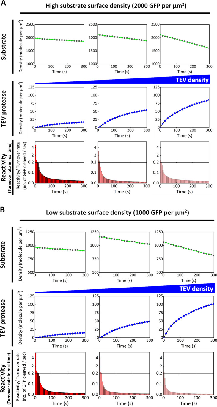
The cell membrane is a critical component of cellular architecture, serving not only as a physical barrier enclosing the cytosol but also as a dynamic platform for various biochemical reactions. Due to the unique two-dimensional and fluidic environment of the membrane, reactions that occur on its surface are subject to specific physical constraints. While membrane-mediated reactions are known to play key roles in cellular regulation, their advantages and limitations remain inadequately explored. In this study, we reconstitute a classic proteolytic cleavage reaction at the membrane interface, designed for the real-time kinetic analysis down to the single-molecule level. By systematically altering the enzyme-membrane affinity, we examined enzyme-substrate interactions under various conditions. Our findings reveal that while the membrane environment significantly enhances enzymatic turnover rate, it also imposes diffusion limitations that immediately reduce this turnover rate over time. By adjusting the enzyme's membrane affinity to an intermediate level, we enable the enzyme to "hop" on the membrane surface, overcoming these diffusion constraints and sustaining high enzymatic turnover rate with faster kinetics. These results highlight the dual role of the membrane environment in regulating biochemical reactions, balancing enhanced reactivity with physical limitations. Moreover, the ability to dynamically tune membrane affinity to optimize reactions underscores the cell's capacity to regulate enzymatic processes efficiently. This study provides critical insights into the role of the cell membrane in biochemical reactions and offers a broadly applicable framework for understanding membrane-associated interactions in biological systems.


Figures

1
1
(A) Comparison of TEV proteolytic reactions in different scenarios. (B) Schematic of the TEV proteolytic reaction reconstituted on the SLB. His-tagged GFP is anchored to the SLB containing 8% Ni-NTA DOGS. A TEV cleavage site is positioned between the His-tag and GFP. His-tagged TEV is recruited to the SLB via His-tag chemistry and undergoes lateral diffusion. TEV recognizes and cleaves the substrate at the cleavage site, releasing GFP from the SLB. The enzyme turnover rate (corresponding to real-time reactivity) is determined by monitoring the decrease in GFP fluorescence on the membrane surface. (C) Modulation of His4-TEV affinity to the membrane by adjusting the interaction between the His-tag and Ni-NTA lipids. (D) Model of the hopping mechanism. After recruitment to the membrane, TEV undergoes 2D diffusion and reacts with nearby substrates. As local substrate depletion occurs, further cleavage is limited by the slow diffusion of substrates on the SLB. Imidazole can displace His4-TEV from Ni-NTA lipids, releasing TEV into solution, where it undergoes faster 3D diffusion and is recruited back to regions with higher substrate density. Repeated cycles of this hopping mechanism enable TEV to overcome local substrate depletion and extend the range of the proteolytic reaction.
2
2
Enzymatic reaction facilitated initially by the 2D surface through higher local concentration. (A) TEV proteolytic cleavage reaction on the SLB. Fluorescently labeled His7-TEV is recruited to the SLB (blue dots). GFP anchored on the membrane is released from the surface due to the proteolytic cleavage reaction (black dots). (B) TEV proteolytic cleavage reaction between 2D and 3D systems. Cleavage occurs when TEV in solution collides with the substrate anchored on the SLB. The number of non-His-tagged TEV molecules distributed within a 100 nm-thick volume above the SLB is calculated to be ∼9000 in the imaging area, based on the concentration in the bulk solution. (C) TEV proteolytic cleavage reaction in solution. TEV (final concentration: 1 μM) is mixed with GFP (final concentration: 50 μM) in 0.1 M PB buffer. SDS-PAGE is used to track the kinetics of the cleavage reaction. Enzyme turnover rate is determined by calculating the ratio of cleaved to uncleaved substrate band intensities using ImageJ. A visual break in the y-axis is used between 0.2 and 0.5 GFP molecules cleaved per second to accommodate the data range.
3
3
Turnover rate of TEV in real time depends on its surface density. During the reaction, the number of GFP molecules on the SLB decreases due to proteolytic cleavage by TEV (black dot). After the injection of His7-TEV into the sample chamber, His7-TEV is recruited to the membrane via His-tag chemistry (blue dot). The time course of TEV turnover rate is shown by the red bar diagram. Each bar indicates the average number of substrate molecules cleaved per second by one TEV protease. The real-time turnover rate depends on the surface density of the enzyme and reflects competition for substrates among enzymes. The data demonstrate that as more TEV proteases are recruited to the membrane, the real-time turnover rate per enzyme decreases due to the limited number of substrates available to each enzyme. The surface densities of the substrate are controlled at approximately 2000 and 1000 molecules per μm2 in (A) and (B), respectively, and both show similar trends in real-time activity across three different final TEV densities. Statistical analyses of the experiments are provided in Figure S10 and S11.
4
4
Initial turnover rate of TEV is markedly enhanced by the 2D environment of the SLB when TEV is at the single-molecule level and substrates are in large excess at the start of the reaction. The initial turnover rate increases significantly when both TEV and its substrates are anchored to the SLB and substrate competition among enzymes is minimal during the initial phase of the reaction. The surface density of GFP-tagged substrates on the SLB is controlled at 5000 molecules per μm2 (black dot), while the number of TEV molecules recruited to the membrane is controlled at 400–500 (a surface density of approximately 0.12 molecules per μm2) within the imaging field (blue dot). The initial turnover rate of TEV exceeds 100 substrate molecules per second (red bar). Statistical analyses of the experiments are provided in Figure S10 and S11.
5
5
Modulation of His4-TEV dwell time on the membrane by varying the concentration of imidazole. The distribution of the dwell time for His4-TEV on the membrane is measured at four different concentrations of imidazole, shown in black (0 mM), cyan (10 mM), red (20 mM), and blue (40 mM). Imidazole competes with the His-tag for binding to the Ni-NTA lipid, causing the dissociation of His4-TEV into the solution above and shortening its dwell time on the membrane. The histogram shows that the dwell time of His4-TEV greater than 0.5 s is highly populated when there is no imidazole in the system, while the distribution of dwell times shifts to the short time scale (<0.5 s) as the concentration of imidazole increases.
6
6
Hopping mechanism allows the enzyme to escape from localized movement due to short dwell times on the membrane, unlike molecules that remain permanently anchored. mCherry is anchored to the membrane via maleimide chemistry, with a TEV cutting site between the introduced cysteine at the N-terminus and mCherry. The surface density of mCherry is controlled at about 350 molecules per μm2. The type of movement on the membrane is changed from 2D diffusion to the hopping model by modulating the affinity between the His-tag and Ni-NTA lipid through the addition of imidazole. TEV will eventually collide with the membrane from solution when the imidazole concentration is high enough. (A) Without imidazole ([imidazole] = 0 mM) in the system, the interaction between the His-tag and Ni-NTA lipid is strong. His4-TEV is recruited to the membrane via His-tag chemistry and undergoes 2D diffusion on the membrane. The initial TEV turnover rate is enhanced by the 2D surface (to about 0.7 substrate molecules cleaved per second per TEV), but it soon drops rapidly due to diffusion-limited conditions between enzymes and substrates on the membrane. (B) A low concentration of imidazole ([imidazole] = 40 mM) reduces the affinity between His4-TEV and the SLB containing 4% Ni-NTA lipid. This moderate affinity results in the hopping movement of TEV on the SLB. Under this condition, TEV undergoes cycles of recruitment to the SLB, 2D diffusion, and dissociation from the SLB, effectively expanding the range of the proteolytic cleavage reaction by a single TEV molecule. The leveling off of TEV density reflects equilibrium between its dissociation from and recruitment to the membrane. TEV can thus maintain a relatively high turnover rate by avoiding local substrate depletion (left panel). However, in the presence of a higher density of TEV, the turnover rate drops quickly due to rapid substrate depletion. The same experiments using a different fluorescent protein, mScarlet, are shown in Figure S9. (C) His4-TEV is no longer recruited to the membrane at high imidazole concentrations ([imidazole] = 200 mM). In this case, TEV reacts with the substrate only through collisions from solution, resulting in a relatively low turnover rate. A surface density of TEV comparable to the hopping condition in (B) is achieved by adjusting the concentration of protein in the sample injection. Statistical analyses of the experiments are provided in Figures S10.

Similar articles

References

    1. Sharon N., Lis H.. Lectins as cell recognition molecules. Science. 1989;246:227–234. doi: 10.1126/science.2552581. - DOI - PubMed
    1. Kholodenko B. N., Hoek J. B., Westerhoff H. V.. Why cytoplasmic signalling proteins should be recruited to cell membranes. Trends Cell Biol. 2000;10:173–178. doi: 10.1016/S0962-8924(00)01741-4. - DOI - PubMed
    1. Leo A., Schraven B.. Adapters in lymphocyte signalling. Curr. Opin Immunol. 2001;13:307–316. doi: 10.1016/S0952-7915(00)00220-X. - DOI - PubMed
    1. Malissen B., Grégoire C., Malissen M., Roncagalli R.. Integrative biology of T cell activation. Nat. Immunol. 2014;15:790–797. doi: 10.1038/ni.2959. - DOI - PubMed
    1. Lim, W. ; Mayer, B. ; Pawson, T. . Cell Signaling: Principles and Mechanisms; Garland Science, 2015.

LinkOut - more resources