. 2025 Jun 24;20(6):e0326457.
doi: 10.1371/journal.pone.0326457. eCollection 2025.

Non-invasive tape sampling of tryptophan and kynurenine in relation to phenylalanine and tyrosine from melanoma and adjacent non-lesional skin: A pilot study

Affiliations

Non-invasive tape sampling of tryptophan and kynurenine in relation to phenylalanine and tyrosine from melanoma and adjacent non-lesional skin: A pilot study

Skaidre Jankovskaja et al. PLoS One. .

Abstract

Purpose: To evade immunosurveillance many cancers convert tryptophan (Trp) into kynurenine (Kyn), which induces immunotolerance and suppresses immune responses. Elevated Kyn amounts have been found in blood from patients with cutaneous melanoma. This study aimed to investigate whether higher Kyn abundance and lower Trp abundance can be detected on the surface of cutaneous melanoma lesions compared with adjacent non-lesional skin.

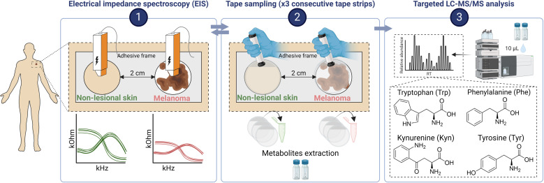
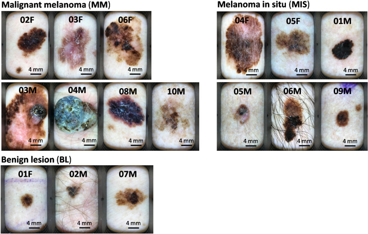
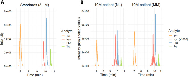
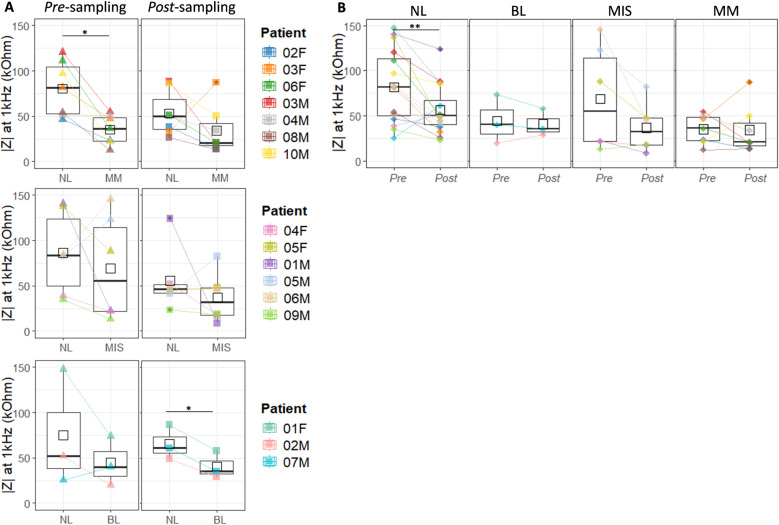
Methods: Sixteen patients with suspected melanomas were enrolled in this study. All lesions were excised and histopathologically diagnosed: 7 lesions were diagnosed as invasive malignant melanomas (MM), 6 as melanomas in situ (MIS), and 3 as benign lesions (BL). Non-invasive metabolite sampling was performed by tape stripping of suspected skin lesions and adjacent healthy non-lesional (NL) skin. Trp, Kyn, tyrosine (Tyr), and phenylalanine (Phe) were quantified by liquid chromatography-tandem mass spectrometry (LC-MS/MS). Electrical impedance spectroscopy (EIS) measurements were conducted to assess skin barrier integrity.

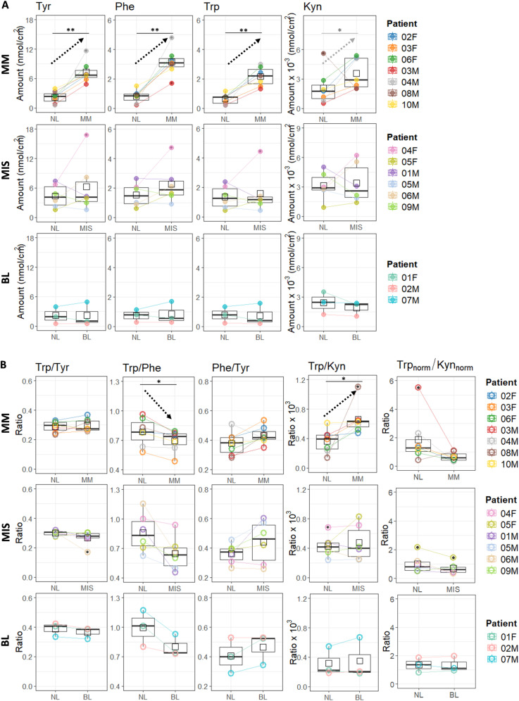
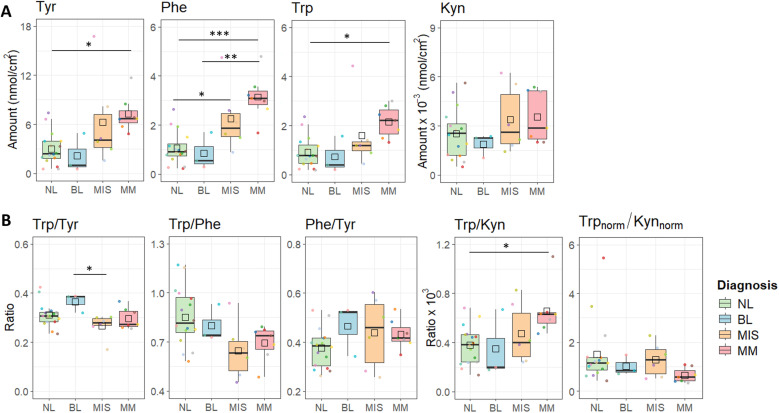
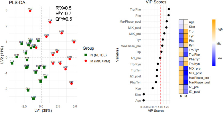
Results: Levels of all metabolites, Tyr (x6), Phe (x6), Trp (x5), and Kyn (x3), were significantly higher in MM lesions compared with adjacent NL skin, resulting in an elevated Trp/Kyn ratio. Trp levels increased less than Phe and Tyr levels, suggesting a potential increase in Trp depletion. Skin resistance in MM lesions was half that of NL skin. No differences were observed between MIS or BL and NL skin.

Conclusions: Non-invasive skin sampling revealed elevated Tyr, Phe, Trp and Kyn levels in MM skin, which is likely the result of compromised skin barrier at this stage of cutaneous melanoma.


Conflict of interest statement

The authors have declared no competing interests exist.

Figures

Fig 1
Fig 1. Experimental design of the study performed on melanoma-suspected study participants.
A frame, designed to minimize the sampling of non-lesional skin along with melanoma-suspected lesion, was fixed on the skin, separating the two sampling areas by 2 cm. EIS measurements were conducted on both sampling areas prior tape sampling. Then, adhesive sampling with tapes was performed three times on each site with a new tape each time. After that, post sampling EIS measurements were conducted on the same lesions. LMW compounds were then extracted from the tapes and the abundance of four amino acids was determined by LC-MS/MS. Created with BioRender.com.
Fig 2
Fig 2. Dermatoscopic images of melanoma-suspected skin lesions.
The study participants indicated on the top of the image correspond to patient names in Table 1.
Fig 3
Fig 3. LC-MS/MS chromatograms of a standard solution (A) and a patient sample (B).
Fig 4
Fig 4. The quantities of Tyr, Phe, Trp and Kyn (A), and their ratios (B), tape sampled from the surface of non-lesional (NL) skin compared with malignant melanoma (MM, n = 7), melanoma in situ (MIS, n = 6), and benign lesions (BL, n = 3).
Trpnorm = Trp/Phe and Kynnorm = Kyn/Kynaverage. Comparison of melanoma-suspected skin lesions with non-lesional (NL) skin, was done by using paired two-sample t-test followed by FDR correction. Significance levels: ‘*’ p < 0.05, ‘**’ p < 0.01.
Fig 5
Fig 5. Comparative analysis of metabolite quantities (A) and their ratios (B) across skin surface samples (NL, BL, MIS, MM).
Metabolite quantities and their ratios were compared across different skin surface samples using one-way ANOVA followed by a post-hoc Tukey test. Significance levels are indicated as: ‘*’ p < 0.05, ‘**’ p < 0.01, ‘***’ p < 0.001.
Fig 6
Fig 6. Evaluation of skin barrier integrity using impedance at 1 kHz (skin resistance).
(A) Comparison of skin resistance between BL, MIS, MM lesions, and adjacent NL skin, measured before (pre-sampling) and after (post-sampling) tape sampling. (B) Effect of tape stripping on skin resistance, comparing measurements taken before (Pre) and after (Post) tape sampling. Comparison between two sample groups was done by using paired two-sample t-test test followed by FDR correction. Significance levels: ‘*’ p < 0.05, ‘**’ p < 0.01.
Fig 7
Fig 7. Partial least squares-discriminant analysis (PLS-DA) for two patient groups: non-melanoma (N, including NL and BL samples) and melanoma (M, including MIS and MM samples).
Variables were ranked by variable importance projection (VIP) scores, where VIP scores > 1 reflect most important variables in separating between non-melanoma vs. melanoma sample groups. The heat map shows how variable levels (e.g., metabolite abundance, impedance values, lesion size) contribute to N and M groups separation.

Similar articles

References

    1. Bray F, Laversanne M, Sung H, Ferlay J, Siegel RL, Soerjomataram I, et al. Global cancer statistics 2022: GLOBOCAN estimates of incidence and mortality worldwide for 36 cancers in 185 countries. CA Cancer J Clin. 2024;74(3):229–63. doi: 10.3322/caac.21834 - DOI - PubMed
    1. World Health Organization (WHO). Ultraviolet (UV) radiation and skin cancer. 2017. [cited 17 Feb 2025]. https://www.who.int/news-room/questions-and-answers/item/radiation-ultra...
    1. Arnold M, Singh D, Laversanne M, Vignat J, Vaccarella S, Meheus F, et al. Global Burden of Cutaneous Melanoma in 2020 and Projections to 2040. JAMA Dermatol. 2022;158(5):495–503. doi: 10.1001/jamadermatol.2022.0160 - DOI - PMC - PubMed
    1. Zbytek B, Carlson JA, Granese J, Ross J, Mihm MC Jr, Slominski A. Current concepts of metastasis in melanoma. Expert Rev Dermatol. 2008;3(5):569–85. doi: 10.1586/17469872.3.5.569 - DOI - PMC - PubMed
    1. Balch CM, Gershenwald JE, Soong S-J, Thompson JF, Atkins MB, Byrd DR, et al. Final version of 2009 AJCC melanoma staging and classification. J Clin Oncol. 2009;27(36):6199–206. doi: 10.1200/JCO.2009.23.4799 - DOI - PMC - PubMed

MeSH terms