. 2025 Jun 24;20(6):e0317387.
doi: 10.1371/journal.pone.0317387. eCollection 2025.

Evaluation of subretinally delivered Cas9 ribonucleoproteins in murine and porcine animal models highlights key considerations for therapeutic translation of genetic medicines

Affiliations

Evaluation of subretinally delivered Cas9 ribonucleoproteins in murine and porcine animal models highlights key considerations for therapeutic translation of genetic medicines

Spencer C Wei et al. PLoS One. .

Abstract

Genetic medicines, including CRISPR/Cas technologies, extend tremendous promise for addressing unmet medical need in inherited retinal disorders and other indications; however, there remain challenges for the development of therapeutics. Herein, we evaluate genome editing by engineered Cas9 ribonucleoproteins (eRNP) in vivo via subretinal administration using mouse and pig animal models. Subretinal administration of adenine base editor and double strand break-inducing Cas9 nuclease eRNPs mediate genome editing in both species. Editing occurs in retinal pigmented epithelium (RPE) and photoreceptor cells, with favorable tolerability in both species. Using transgenic reporter strains, we determine that editing primarily occurs close to the site of administration, within the bleb region associated with subretinal injection. Our results show that subretinal administration of BE-eRNPs in mice mediates base editing of up to 12% of the total neural retina, with an average rate of 7% observed at the highest dose tested. In contrast, a substantially lower editing efficiency was observed in minipigs; even with direct quantification of only the treated region, a maximum base editing rate of 1.5%, with an average rate of <1%, was observed. Our data highlight the importance of species consideration in preclinical studies for the development of genetic medicines targeting the eye and provide an example of a lack of translation between small and larger animal models in the context of subretinal administration of Cas9 eRNPs.


Conflict of interest statement

All authors except PKS, BRP, DG, and KS are employees of Spotlight Therapeutics, Inc., or were employees at the time the research took place. RM, KM, SCW, BGG, and MJJ are inventors on patent applications related to delivery of Cas9 ribonucleoproteins (PCT/US2020/024249 and PCT/US2023/076149). SCW is a prior consultant for BioEntre, consultant for Actym Therapeutics, an inventor on a patent for a mouse model of autoimmune adverse events (PCT/US2019/050551), and employee of Inversion Therapeutics. Benjamin Gowen is an employee of Editpep, Inc. KS is a member of the scientific advisory boards of Andson Biotech, Bharat Biotech, and Notch Therapeutics and is a board member for Andson Biotech and Bharat Biotech. This does not alter our adherence to the PLOS ONE policies on sharing data and materials.

Figures

Fig 1
Fig 1. Overview of engineered ribonucleoprotein (eRNP) approach and challenges to treat inherited retinal disease.
A. Features of adenine base editing engineered ribonucleoprotein (BE-eRNP). CPP, cell-penetrating peptide; sgRNA, single guide RNA. B. Subretinal delivery route and retinal anatomy. The bleb is formed between the outer nuclear layer containing photoreceptors and the retinal pigmented epithelium. Key landmark anatomical structures are labeled. C. Aligned genomic DNA sequences from the indicated species and alleles corresponding to one human disease variant underlying Stargardt’s Disease, ABCA4 c.5882G > A, p.G1961E. The sequences shown correspond to a protospacer sequence and protospacer adjacent motif (PAM, yellow highlight) for WT SpCas9 with the disease-causing missense mutation (A in the human disease allele) colored red. A bystander A, colored blue, is present in all species shown but would not change the coding sequence of the gene if edited to G by an ABE. Lowercase bases indicate positions in the protospacer that vary relative to the human sequences.
Fig 2
Fig 2. Identification of high-efficiency ABE gRNAs targeting the mouse Rosa26 locus and eRNP editing upon subretinal injection in mice.
A. Editing rates in mouse T cells for different gRNAs delivered by ABE-eRNPs. ABE-eRNPs were complexed with the indicated gRNAs in crRNA:trcrRNA format and nucleofected into cells. Editing rates were quantified based on read frequency from Illumina sequencing and are displayed the frequency of reads for the highest frequency sequence among all sequences with any A-to-G edit in the 3–8 base pair edit window from the 5’ end of each respective protospacer sequence.. The asterisk indicates the crRNA corresponding to the targeting gRNA used in panels (B) and (C). B. Editing rates by tissue following subretinal injection of Rosa26-targeting ABE-eRNPs complexed with mmRosa_sg1 in mice. 7 days following subretinal administration, mice were sacrificed and their eyes were dissected to separate the neural retina from the eyecup (containing choroid, sclera and RPE). Editing rates from Illumina DNA amplicon sequencing are shown as mean ± standard deviation (SD) at each dose. Each overlaid point corresponds to a single eye. Reads were scored as positive for editing if at least one A → G transition was detected within a 10-base editing window. C. Editing rates from the same samples in (B) plotted as editing in retina vs. eyecup tissues, colored by RNP dose. Each point corresponds to one eye. D. Fluorescent images of entire RPE tissue florets from Ai14 mice dissected from enucleated eyes 2 weeks after injection with NHEJ-eRNP complexed with sgAi14. The RPE florets were flat-mounted to assess genome editing by imaging the tdTomato (red) fluorescence through confocal microscopy.
Fig 3
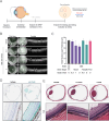
Fig 3. Physiology and histology of porcine eyes following subretinal administration.
A. Graphical summary of the experimental design to evaluate the effects of subretinal administration of eRNP in Yucatan minipigs. B. OCT images of porcine eyes immediately following and 2 weeks following subretinal dosing of vehicle or ABE-eRNPs at the indicated dosages (b-scan indicated by green arrow). C. Outer nuclear layer thickness according to OCT at 2 weeks post injection plotted for ABE-eRNP groups. Data are plotted as the mean ± SD for each group across all measurement areas from superior (injected) regions, as shown for representative eyes in (A). D. Representative images of porcine eyes 4 hours post subretinal administration stained with isotype control or anti-Cas9 immunohistochemistry (teal) at 2.5× (top) and 20× (bottom) magnification. E. Representative whole eye scans and high magnification (inset) images of H&E-stained porcine eyes 2 weeks post subretinal administration with either vehicle, 0.3 nmol ABE-eRNPl or 3 nmol ABE-eRNP.
Fig 4
Fig 4. Adenine base editing following ABE-eRNP subretinal administration in minipig eyes.
A. Editing rates by tissue following subretinal injection of ROSA26-targeting ABE-eRNPs complexed with sgRosa26 in minipigs at multiple doses. Two weeks following subretinal administration, animals were sacrificed, and eyes were dissected to collect intact tissue layers for the neural retina and the choroid + retinal pigmented epithelium (RPE). For each tissue, three biopsy punches were taken corresponding to the injection bleb, tissue immediately adjacent to the bleb, and a region distal to the bleb. Editing rates from Illumina DNA amplicon sequencing are plotted for each eye by tissue, biopsy region, and dose. Reads were scored as positive for editing if at least one A → G transition was detected within a 10 base edit window. Gray lines connect points corresponding to one eye. B. Editing rates as shown in (A) for ABE-eRNPs complexed with sgABCA4.
Fig 5
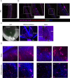
Fig 5. Characterization of editing in the transgenic SRM1 pig strain.
Pseudocolored images show staining by DAPI (blue), native tdTomato (red), anti-tdTomato-Cy5 (magenta) A. Fluorescent image of cryosection of an SRM1 transgenic porcine eye treated with a negative control non-targeting gRNA NHEJ-eRNP. RPE65 is co-stained for RPE visualization (green). B. Fluorescent images of the superior (treated) region of the two SRM1 transgenic porcine eyes subretinally administered targeting gRNA NHEJ-eRNP. Scale bar, 50 microns. Insets show higher magnification of the boxed regions. C. OCT (left) and fluorescent images of flat-mounted tissue (middle and right) after subretinal administration of eRNP. The bleb region is outlined with a dotted line with anatomical regions labeled. D. Fluorescent images of retina and RPE/choroid/sclera flat-mounted tissue for eyes from the two SRM1 pigs administered targeting gRNA NHEJ-eRNP. Insets show higher magnification of the boxed regions.

Update of

References

    1. Macarrón Palacios A, Korus P, Wilkens BGC, Heshmatpour N, Patnaik SR. Revolutionizing in vivo therapy with CRISPR/Cas genome editing: breakthroughs, opportunities and challenges. Front Genome Ed. 2024;6. doi: 10.3389/fgeed.2024.1342193 - DOI - PMC - PubMed
    1. Raguram A, Banskota S, Liu DR, 2022. Therapeutic in vivo delivery of gene editing agents. Cell 185, 2806–27. doi: 10.1016/j.cell.2022.03.045 - DOI - PMC - PubMed
    1. Taha EA, Lee J, Hotta A. Delivery of CRISPR-Cas tools for in vivo genome editing therapy: trends and challenges. J Control Release. 2022;342:345–61. doi: 10.1016/j.jconrel.2022.01.013 - DOI - PubMed
    1. Musunuru K, Grandinette SA, Wang X, Hudson TR, Briseno K, Berry AM, et al. Patient-specific in vivo gene editing to treat a rare genetic disease. N Engl J Med. 2025;392(22):2235–43. doi: 10.1056/NEJMoa2504747 - DOI - PubMed
    1. Cong L, Ran FA, Cox D, Lin S, Barretto R, Habib N, et al. Multiplex genome engineering using CRISPR/Cas systems. Science. 2013;339(6121):819–23. doi: 10.1126/science.1231143 - DOI - PMC - PubMed

Substances