. 2025 Jun 24;16(1):5331.
doi: 10.1038/s41467-025-60830-1.

Apolipoprotein M attenuates age-related macular degeneration phenotypes via sphingosine-1-phosphate signaling and lysosomal lipid catabolism

Affiliations

Apolipoprotein M attenuates age-related macular degeneration phenotypes via sphingosine-1-phosphate signaling and lysosomal lipid catabolism

Tae Jun Lee et al. Nat Commun. .

Abstract

Age-related macular degeneration (AMD) is a leading cause of blindness in people over 50. AMD and cardiovascular disease share risk factors including age, impaired lipid metabolism, and extracellular lipid deposition. Because of its importance in age-related diseases, we hypothesize that apolipoprotein M (ApoM), a lipocalin that binds sphingosine-1-phosphate (S1P), might restore lipid homeostasis and retinal function in AMD. In support, we find that human patients with AMD demonstrate significantly reduced ApoM compared to controls. In mice with impaired retinal cholesterol efflux, ApoM improves retinal pigment epithelium (RPE) function and lipotoxicity in an S1P- and S1P receptor 3-dependent manner. Ultrastructural evidence of enhanced melanosome-lipid droplet interactions led us to hypothesize and demonstrate that ApoM-S1P signaling drives RPE-specific lysosomal lipid catabolism. RPE-specific knockout of lysosomal acid lipase recapitulates features of AMD. Our study defines a novel role for ApoM/S1P signaling in AMD driven by RPE lipotoxicity, mediated by cell-autonomous lysosomal lipid catabolism.


Conflict of interest statement

Competing interests: RSA and AJ have intellectual property applications licensed by Washington University to Mobius Scientific. RSA and AJ are on the advisory board of Mobius Scientific, and RSA is currently the CSO at Mobius Scientific. The remaining authors declare no competing interests.

Figures
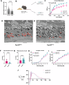
Fig. 1
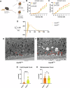
Fig. 1. ApoM therapy ameliorates a dry AMD-like phenotype.
A ApoM levels controlled to total cholesterol levels were measured in patients with (n = 53) or without AMD (n = 328) aged 65 and over. Patients with AMD had significantly lower ApoM levels compared to those without AMD (****p = 0.0000000006, two-tailed Welch’s t-test). B High fat diet fed Abca1/g1-rod/-rod mice were treated with either ApoMKO or ApoMTG plasma (created in BioRender. Lee, T. (2025) https://BioRender.com/li4w1zf). C RPE functional assessment using light bleach dark adaptation electroretinography (n = 6) revealed significantly improved functional recovery in ApoMTG plasma treated mice (*p = 0.0271, Two-way ANOVA). D, E Representative TEM imaging of RPE from either ApoMKO or ApoMTG plasma treated Abca1/g1-rod/-rod mice. Red arrows point to lipid droplets. F Quantitative assessment of lipid droplets per high-powered field within the RPE (n = 30) revealed significantly reduced lipid droplets in ApoMTG plasma treated mice (****p = 0.0000011, two-tailed Welch’s t-test). G Quantitative assessment of melanosomes per high-powered field within the RPE (n = 30) revealed significantly increased melanosome counts in ApoMTG plasma treated mice (****p = 0.0000068, two-tailed Welch’s t-test). Full-field scotopic electroretinography (n = 6) demonstrated improved H rod photoreceptor (*p = 0.0343, Two-way ANOVA) but not I inner retina (Two-way ANOVA) function in ApoMTG plasma-treated mice. J Representative traces of 0 dB intensity flashes show an improved waveform from ApoMTg+ injected mice. The scale bar represents 2 μm. Values are mean ± SE. Source data are provided as a Source Data file.
Fig. 2
Fig. 2. Binding of S1P by ApoM is necessary for RPE lipotoxicity amelioration.
A High fat diet fed Abca1/g1-rod/-rod were treated with plasma from either control mice with human ApoM knocked in (ApoMCtrl) or mice expressing ApoM incapable of binding S1P (ApoMTM) (created in BioRender. Lee, T. (2025) https://BioRender.com/li4w1zf). Full-field scotopic electroretinography (n = 4) demonstrated significant reduction in B rod photoreceptor (*p = 0.0469, Two-way ANOVA) and C inner retina function (*p < 0.0199, Two-way ANOVA) in ApoMTM treated mice. D Representative traces of 0 dB intensity flashes show the waveform from ApoMTM treated mice do not recover. E, F Representative TEM imaging of RPE from either ApoMCtrl or ApoMTM plasma treated Abca1/g1-rod/-rod mice. Red arrows point to lipid droplets. G Quantitative assessment of lipid droplets per high-powered field within the RPE (n = 45) revealed significantly increased lipid droplets in ApoMTM plasma treated mice (****p < 0.000000013, two-tailed Welch’s t-test). H Quantitative assessment of melanosomes per high-powered field within the RPE (n = 45) revealed no significant difference according to two-tailed Welch’s t-test without multiple comparisons in melanosome counts between ApoMCtrl and ApoMTM plasma treated mice. Scale bar represents 2 μm. Values are mean ± SE. Source data are provided as a Source Data file.
Fig. 3
Fig. 3. S1pr3 knockout is sufficient to provoke a dry AMD-like RPE phenotype.
A S1pr3 germline knockout mice (S1pr3−/−) were generated and placed on 6 weeks of high fat diet and subsequently assessed (created in BioRender. Lee, T. (2025) https://BioRender.com/li4w1zf). B RPE functional assessment using light bleach dark adaptation electroretinography (n = 10) revealed significantly reduced functional recovery in S1pr3−/− mice compared to wild-type mice (*p = 0.0462, Two-way ANOVA). C, D Representative TEM imaging of RPE from either wild-type or S1pr3 knockout mice. Red arrows point to lipid droplets. E Quantitative assessment of lipid droplets per high-powered field within the RPE (n = 80) revealed significantly increased lipid droplets in S1pr3 knockout mice (****p = 0.0000000001, two-tailed Welch’s t-test). F Quantitative assessment of melanosomes per high-powered field within the RPE (n = 80) revealed significantly decreased melanosomes in S1pr3 knockout mice (**p = 0.0018, two-tailed Welch’s t-test). Full-field scotopic electroretinography (n = 8) demonstrated reduced G rod photoreceptor (**p = 0.0018, Two-way ANOVA) but not H inner retina (Two-way ANOVA) function in S1pr3 knockout mice. I Representative traces of 0 dB intensity flashes show a diminutive waveform from S1pr3−/−. Scale bar represents 2 μm. Values are mean ± SE. Source data are provided as a Source Data file.
Fig. 4
Fig. 4. ApoM overexpression does not rescue S1PR3 deficiency-induced RPE lipotoxicity.
A S1pr3 germline knockout mice (S1pr3−/−) were crossed with ApoM overexpressing transgenic mice (ApoMTG) to generate S1pr3−/−/ApoMTG mice and placed on 6 weeks of high fat diet and subsequently assessed (created in BioRender. Lee, T. (2025) https://BioRender.com/li4w1zf). B RPE functional assessment using light bleach dark adaptation electroretinography (n = 6) revealed no significant differences in functional recovery between S1pr3−/− and S1pr3−/−/ApoMTG mice. C, D Representative TEM imaging of RPE from either S1pr3−/− or S1pr3−/−/ApoMTG mice. Red arrows point to lipid droplets. E Quantitative assessment of lipid droplets per high-powered field within the RPE (n = 42) revealed no significant differences using two-tailed Welch’s t-test without multiple comparisons between S1pr3−/− and S1pr3−/−/ApoMTG mice. Full-field scotopic electroretinography (n = 8) demonstrated no significant differences by two-way ANOVA in F rod photoreceptor and G inner retina between S1pr3−/− and S1pr3−/−/ApoMTG mice. H Representative traces of 0 dB intensity flashes show waveforms do not improve despite ApoMTG treatment in S1pr3−/−. Scale bar represents 2 μm. Values are mean ± SE. Source data is provided as a Source Data file.
Fig. 5
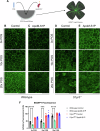
Fig. 5. RPE from ex vivo eyecups recapitulate in vivo lipid burden.
A The RPE/choroid/sclera complex was isolated from mouse eyecups to assess ex vivo RPE for lipid burden using BODIPY neutral lipid staining depicted in green (created in BioRender. Lee, T. (2025) https://BioRender.com/li4w1zf). Eyecups from wild-type mice were treated with increasing dosages of photoreceptor outer segments (POS) before being treated with either B control media or C media with ApoM-S1P. The same paradigm was tested in eyecups from S1pr3−/− mice also treated with either D control media or E media with ApoM-S1P. F BODIPY fluorescence was quantified from each group and plotted (n = 8). Wild-type RPE treated with ApoM-S1P expressed significantly less BODIPY fluorescence compared to those treated with control media in 0x (****p = 0.000022, post hoc Bonferroni’s multiple comparisons test), 10x (***p = 0.00017, post hoc Bonferroni’s multiple comparisons test), and 20x POS (****p = 0.0000000001, post hoc Bonferroni’s multiple comparisons test) treated groups (****p = 0.0000000001, Two-way ANOVA). ApoM-S1P treated S1pr3KO RPE did not demonstrate significant differences in BODIPY fluorescence compared to control-treated S1pr3KO RPE. The scale bar represents 100 μm. Values are mean ± SE. Source data is provided as a Source Data file.
Fig. 6
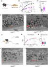
Fig. 6. RPE-specific knockout of lipophagy recapitulate features of a dry AMD-like phenotype.
A RPE-specific lysosomal acid lipase conditional knockout mice (Lipa-RPE/-RPE) were generated and placed on 6 weeks of high fat diet and subsequently assessed (created in BioRender. Lee, T. (2025) https://BioRender.com/li4w1zf). B RPE functional assessment using light bleach dark adaptation electroretinography (n = 6) revealed significantly reduced functional recovery in Lipa-RPE/-RPE mice compared to wild-type mice (***p = 0.000468, Two-way ANOVA). C) Quantitative assessment of lipid droplets per high-powered field within the RPE (n = 76) revealed significantly increased lipid droplets in Lipa-RPE-RPE mice (****p = 0.00000000001, two-tailed Welch’s t-test). D, E Representative TEM imaging of RPE from either wild-type or Lipa-RPE/-RPE mice. Red arrows point to lipid droplets. F High fat diet fed Lipa-RPE/-RPE mice were treated with either ApoMKO or ApoMTG plasma (created in BioRender. Lee, T. (2025) https://BioRender.com/li4w1zf). G RPE functional assessment using light bleach dark adaptation electroretinography (n = 6) revealed no significant differences by two-way ANOVA in functional recovery. H Quantitative assessment of lipid droplets per high-powered field within the RPE (n = 100) revealed no significant differences according to two-tailed Welch’s t-test without multiple comparisons in lipid droplet counts. I, J Representative TEM imaging of RPE from either wild-type or Lipa-RPE/-RPE mice. Red arrows point to lipid droplets. The scale bar represents 2 μm. Values are mean ± SE. Source data is provided as a Source Data file.
Fig. 7
Fig. 7. ApoM-S1P as a potential therapeutic for early dry AMD.
ApoM is carried by HDL particles in systemic circulation and is made available to the RPE through the choriocapillaris. ApoM and its binding partner, S1P, activates S1PR3 on the RPE surface to activate lysosomal lipid catabolism to alleviate RPE lipid burden. Inactive S1PR3 allows aberrant accumulation of intracellular lipids within the RPE, leading to dysfunction and potentially increasing the risk to develop dry AMD features. By supplying the RPE with exogenous ApoM-S1P through plasma transfer, we can reduce lipid burden in the RPE through a S1PR3-dependent mechanism and subsequently reduce the risk of dry AMD pathogenesis (created in BioRender. Lee, T. (2025) https://BioRender.com/li4w1zf).

References

    1. Apte, R. S. Age-related macular degeneration. N. Engl. J. Med385, 539–547 (2021). - PMC - PubMed
    1. Wong, W. L. et al. Global prevalence of age-related macular degeneration and disease burden projection for 2020 and 2040: a systematic review and meta-analysis. Lancet Glob. Health2, e106–e116 (2014). - PubMed
    1. Rein, D. B. et al. Prevalence of age-related macular degeneration in the US in 2019. JAMA Ophthalmol.140, 1202–1208 (2022). - PMC - PubMed
    1. Sene, A. et al. Impaired cholesterol efflux in senescent macrophages promotes age-related macular degeneration. Cell Metab.17, 549–561 (2013). - PMC - PubMed
    1. Curcio, C. A. Antecedents of Soft Drusen, the specific deposits of age-related macular degeneration, in the biology of human Macula. Invest Ophthalmol. Vis. Sci.59, AMD182–AMD194 (2018). - PMC - PubMed

MeSH terms

LinkOut - more resources