Review
. 2025 Jun;57(6):1121-1132.
doi: 10.1038/s12276-025-01470-9. Epub 2025 Jun 24.

Caspases as master regulators of programmed cell death: apoptosis, pyroptosis and beyond

Affiliations
Review

Caspases as master regulators of programmed cell death: apoptosis, pyroptosis and beyond

So Hee Dho et al. Exp Mol Med. 2025 Jun.

Abstract

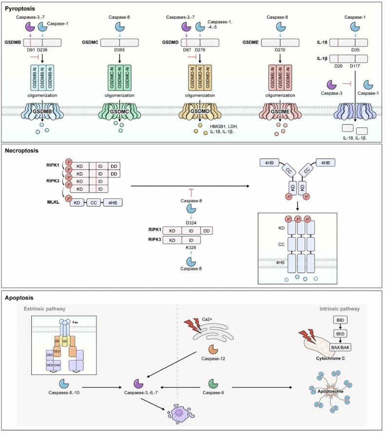
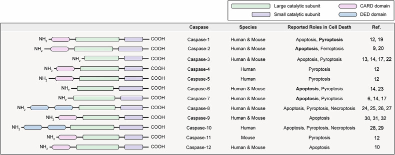
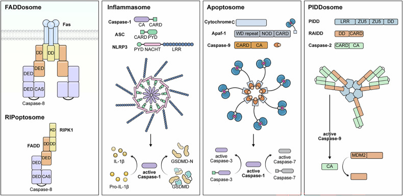
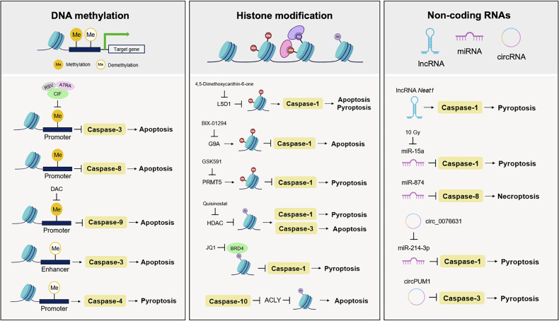
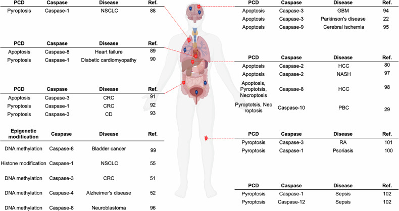
Caspases are crucial regulators of programmed cell death, mediating pathways such as apoptosis, pyroptosis, necroptosis and ferroptosis. Their activity is intricately controlled by epigenetic modifications, molecular interactions and post-translational changes, reflecting their central role in cellular homeostasis and disease mechanisms. Dysregulated caspase functions are linked to a wide array of conditions, including cancer, neurodegenerative disorders and inflammatory diseases, establishing their importance as potential therapeutic targets. The roles and regulation of caspases across subcellular compartments and their molecular interactions provide critical insights into the complexity of programmed cell death. Here, this review synthesizes current knowledge on the diverse functions of caspases, offering a comprehensive foundation for exploring innovative therapeutic strategies.


Conflict of interest statement

Competing interests: The authors declare no competing interests.

Figures

Fig. 1
Fig. 1. Overview of major signaling components of PCD.
The molecular signaling pathways and key effector molecules involved in three major forms of PCD: pyroptosis, necroptosis and apoptosis. Pyroptosis: this process is initiated by the cleavage of GSDM by inflammatory caspases, resulting in the formation of GSDM-N, which creates pores in the plasma membrane and leads to cellular lysis. Simultaneously, pro-inflammatory cytokines such as IL-1β and IL-18 are activated by caspase-1, allowing their release through the GSDM-formed pores. These cytokines amplify immune responses, enhancing the inflammatory reaction to infections or cellular damage. However, caspases also have a regulatory role in inhibiting pyroptosis. For example, caspases-3 and -7 can cleave GSDMD at non-canonical sites, preventing pore formation. Additionally, the cleavage of GSDMB by caspases-3, -6 and -7 can suppress its pore-forming ability. Furthermore, caspase-3 can cleave IL-1β at alternative sites, inhibiting its activation and thereby reducing inflammation. This dual role of caspases in both promoting and suppressing pyroptosis underscores their crucial function in maintaining immune homeostasis. Necroptosis: initiated by the RIPK1/RIPK3-MLKL axis, RIPK3 phosphorylates MLKL, leading to its oligomerization and translocation to the plasma membrane. The incorporation of MLKL into the membrane causes rupture and subsequent cellular death. Necroptosis is tightly regulated to prevent excessive inflammation. Apoptosis: caspase-8 (extrinsic pathway) and caspase-9 (intrinsic pathway) play crucial roles in apoptosis by cleaving and activating executioner caspases, such as caspase-3 and caspase-7. These executioner caspases mediate downstream processes of apoptosis, including the dismantling of cellular structures, which leads to controlled cell death. In the intrinsic pathway, tBID translocates to the mitochondria, activating BAX and BAK, which promote MOMP. This process results in the release of apoptogenic factors from the mitochondria, ultimately leading to cell death during the execution phase of apoptosis. KD RIPK1 kinase domain, ID intermediate domain, DD death domain, CC coiled coil, 4HB 4-helical bundle, DED death effector domain, CAS catalytic subunit.
Fig. 2
Fig. 2. Domain structures and functional roles of caspases in PCD.
The domain structures of caspases, including CARD, DED and the large and small catalytic subunits. Caspases are categorized based on their roles in apoptosis, pyroptosis, necroptosis and ferroptosis. Bold text indicates the primary cell death pathway associated with each caspase. Additionally, the table provides the mouse and human counterparts for each caspase, detailing their expression and functional roles in both species.
Fig. 3
Fig. 3. Regulating cell death through caspase domain interactions.
A schematic illustrating the five major protein complexes involved in PCD pathways—FADDosome, RIPoptosome, inflammasome, apoptosome and PIDDosome. It highlights their composition and roles in activating key caspases and downstream effectors. FADDosome: composed of Fas receptor, FADD and caspase-8. The DD of Fas binds to FADD, which recruits and activates caspase-8 via its DED, thereby initiating extrinsic apoptosis. RIPoptosome: formed by RIPK1, FADD and caspase-8. The RIPK1 kinase domain (KD) and DD facilitate complex formation, leading to caspase-8 activation and subsequent apoptosis, or necroptosis if caspase-8 is inhibited. Inflammasome: a multiprotein platform consisting of NLRP3, ASC and caspase-1. The PYD of NLRP3 interacts with ASC, which in turn recruits caspase-1 via its CARD. Activated caspase-1 processes IL-1β, IL-18 and GSDMD, driving pyroptosis and inflammation. Apoptosome: consists of Apaf-1, cytochrome c and caspase-9. Upon release from the mitochondria, cytochrome c binds Apaf-1, inducing apoptosome assembly. This activates caspase-9, which subsequently activates caspases-3 and -7 to execute intrinsic apoptosis. PIDDosome: composed of PIDD, RAIDD and caspase-2. PIDD recruits RAIDD via DD interactions, which then activates caspase-2 via CARD. Caspase-2 activation modulates apoptosis and DNA damage responses by interacting with substrates such as MDM2.
Fig. 4
Fig. 4. Epigenetic regulation of caspases and its impact on cell death and disease.
Epigenetic regulation occurs through various epigenetic mechanisms, including DNA methylation, histone modification and ncRNAs. First, the methylation status of DNA in promoter and enhancer regions substantially impacts caspase expression. For example, promoter methylation suppresses caspase-3 and caspase-8, thereby inhibiting apoptosis, whereas demethylation of promoter and enhancer regions activates caspase-3 and caspase-9, respectively. Additionally, promoter demethylation facilitates caspase-4-mediated pyroptosis. Second, histone modifications, including methylation and acetylation of histone proteins, regulate chromatin structure and gene transcription. Various histone methyltransferases (for example, LSD1, G9A and PRMT5) modulate histone methylation, influencing caspase-1 activation. HDAC inhibitors, such as Quisinostat, promote caspase-1 activation for pyroptosis and caspase-3 for apoptosis. Furthermore, inhibition of BRD4 by JQ1 induces caspase-1-dependent pyroptosis, while histone acetylation is regulated by caspase-10 through ACLY cleavage. Third, ncRNAs such as lncRNAs, miRNAs and circRNAs also influence caspase activity. For example, lncRNAs such as Neat1 activate caspase-1 to promote pyroptosis, while circRNAs such as circPUM1 activate caspase-3. miRNAs, including miR-15a and miR-874, regulate pyroptosis through caspase-1 and necroptosis through caspase-8. Additionally, circRNAs mediate pyroptosis via the miR-214-3p/caspase-1 pathway. Dysregulation of these epigenetic mechanisms is implicated in numerous diseases, including cancer, neurodegenerative disorders and developmental abnormalities.
Fig. 5
Fig. 5. The role of caspases in PCD pathways and epigenetic regulation across diseases.
The involvement of specific caspases in apoptosis, pyroptosis, necroptosis, histone modification and DNA methylation, and their associations with various diseases, along with their associations with various diseases and the corresponding affected tissues. In apoptosis, caspase-8 is associated with heart failure and HCC, while caspase-3 is linked to GBM, Parkinson’s disease and CRC. Caspase-2 is implicated in NASH and HCC, and caspase-9 is involved in cerebral ischemia. In pyroptosis, caspase-1 contributes to NSCLC, diabetic cardiomyopathy, CRC, psoriasis and sepsis. Caspase-3 is also associated with CD and RA. Additionally, caspase-8 is linked to HCC, while caspase-10 is implicated in PBC. Caspase-12 is involved in sepsis. In the context of necroptosis, caspase-8 plays a notable role in HCC, and caspase-10 is linked to necroptosis in PBC. Regarding histone modification and DNA methylation, caspase-1 mediates histone modification in NSCLC, caspase-8 is associated with DNA methylation in bladder cancer and neuroblastoma, caspase-3 is linked to DNA methylation in CRC and caspase-4 is associated with DNA methylation in Alzheimer’s disease.

Similar articles

References

    1. Van Opdenbosch, N. & Lamkanfi, M. Caspases in cell death, inflammation, and disease. Immunity50, 1352–1364 (2019). - PMC - PubMed
    1. Weindel, C. G., Ellzey, L. M., Martinez, E. L., Watson, R. O. & Patrick, K. L. Gasdermins gone wild: new roles for GSDMs in regulating cellular homeostasis. Trends Cell Biol.33, 773–787 (2023). - PMC - PubMed
    1. Morgan, M. J. & Kim, Y. S. RIPK3 in necroptosis and cancer. Mol. Cells48, 100199–100210 (2025). - PMC - PubMed
    1. Yuan, J., Najafov, A. & Py, B. F. Roles of caspases in necrotic cell death. Cell167, 1693–1704 (2016). - PMC - PubMed
    1. Bertheloot, D., Latz, E. & Franklin, B. S. Necroptosis, pyroptosis and apoptosis: an intricate game of cell death. Cell Mol. Immunol.18, 1106–1121 (2021). - PMC - PubMed

LinkOut - more resources