

Microvesicle inhibition enhances the therapeutic effects of ATRA in acute promyelocytic leukemia cells via changes in miRNAs: the promising antileukemic potential of imipramine
- PMID: 40555813
- PMCID: PMC12187794
- DOI: 10.1007/s10238-025-01763-3
Microvesicle inhibition enhances the therapeutic effects of ATRA in acute promyelocytic leukemia cells via changes in miRNAs: the promising antileukemic potential of imipramine
Abstract
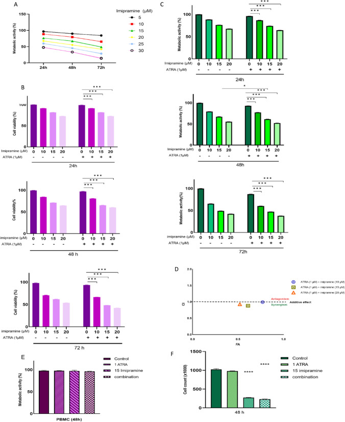
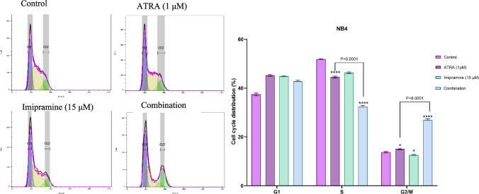
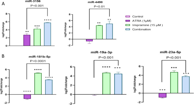
Extracellular vesicles (EVs) represent an essential role in cancer progression through intercellular communication. Therefore, the use of EV formation inhibitors could be a profitable therapeutic strategy in various types of cancer, including leukemia. Imipramine, a tricyclic antidepressant, can block EV formation by inhibiting acid sphingomyelinase. Additionally, other crucial players in cancer progression are microRNAs, which regulate molecular mechanisms at the post-transcriptional level. Here, to potentiate the therapeutic effect of all-trans retinoic acid (ATRA) in acute promyelocytic leukemia (APL), we investigated the effect of imipramine as a microvesicle inhibitor in combination with ATRA for the treatment of APL-derived NB4 cells. Our results declared that imipramine reduced the viability and metabolic activity of ATRA-treated NB4 cells after 48 h. In addition, flow cytometry results highlighted that imipramine induced cytotoxicity through G2/M phase arrest followed by apoptosis. Moreover, we discovered that the antileukemic effects of imipramine were associated with inhibiting microvesicle release and miRNA alteration. Based on bioinformatics methods, we predicted two miRNAs, including hsa-miR-4498 and hsa-miR-3156-5p, which target PML. Additionally, we selected miR-23a-5p, miR-19a-3p, and miR-181b-5p based on relevant studies and subsequently predicted their target genes. The real-time PCR results revealed that the expression level of these miRNAs increased after treatment with imipramine. Moreover, functional enrichment analysis of target genes demonstrated that these genes are involved in cancer-related pathways, including MAPK, FOXO, AMPK, and cellular senescence. Given the significant efficacy of imipramine in potentiating the anti-tumor effects of chemotherapeutic drugs, it can be considered as a potential treatment agent for APL.
Keywords: Bioinformatics; Extracellular vesicle; Imipramine; Leukemia; MicroRNA.
© 2025. The Author(s).
Conflict of interest statement
Declarations. Conflict of interest: The authors declare no competing interests. Consent to publish: Not applicable. Ethical approval: This study was approved by Kerman University of Medical Science Ethical & Research Committee (ethical code: IR.KMU.REC.1400.696). The volunteers received detailed information regarding the aims and methodology of study. Those who expressed willingness to participate, donate blood sample, and sign an informed consent were subsequently recruited. All methods were conducted according to relevant guidelines and applicable regulations.
Figures


References
-
- Arber DA, Orazi A, Hasserjian R, Thiele J, Borowitz MJ, Le Beau MM, et al. The 2016 revision to the World Health Organization classification of myeloid neoplasms and acute leukemia. Blood, J Am Soc Hematol. 2016;127(20):2391–405. - PubMed
MeSH terms
Substances
LinkOut - more resources
Full Text Sources
