. 2025 Jun 25;82(1):255.
doi: 10.1007/s00018-025-05775-w.

miR-29a-3p and TGF-β Axis in Fanconi anemia: mechanisms driving metabolic dysfunction and genome stability

Affiliations

miR-29a-3p and TGF-β Axis in Fanconi anemia: mechanisms driving metabolic dysfunction and genome stability

Nadia Bertola et al. Cell Mol Life Sci. .

Abstract

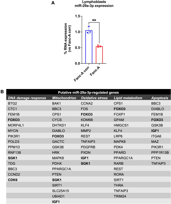
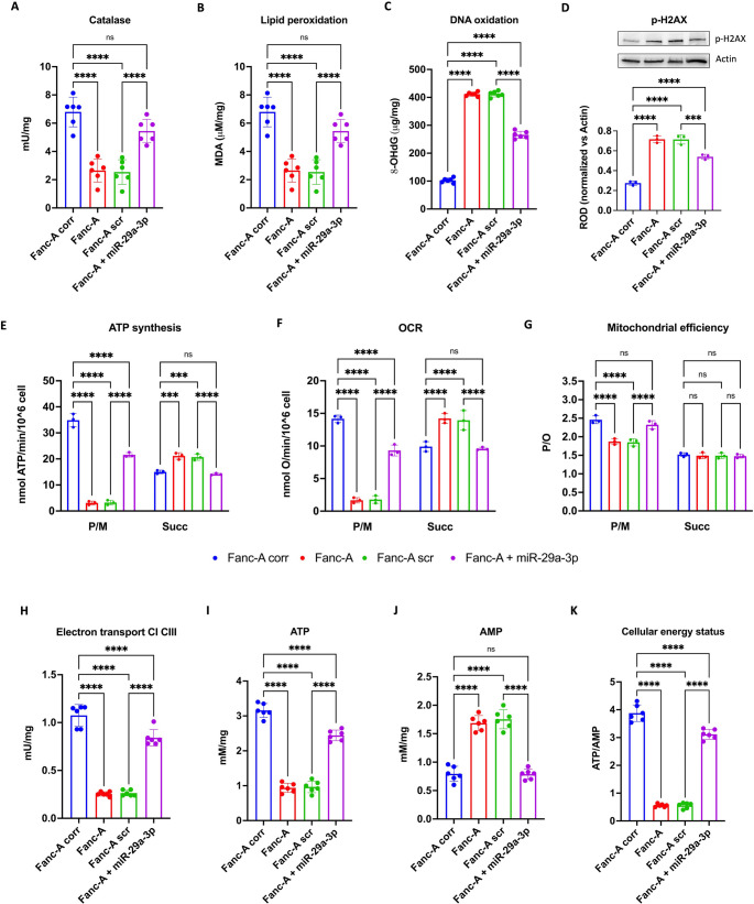
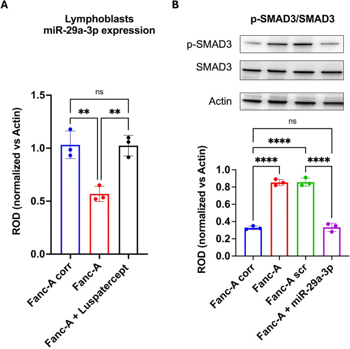
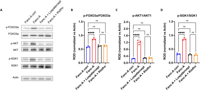
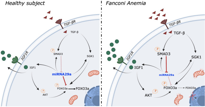
Fanconi anemia (FA) is a genetic disorder characterized by bone marrow failure and cancer predisposition. The FA cellular phenotype is marked by a defective DNA double-strand break repair. Alongside this defect, FA cells exhibit mitochondrial dysfunction and redox unbalance. In addition, FA cells display an altered microRNA profile, including miR-29a-3p, which plays a crucial role in hematopoiesis by supporting the self-renewal, lineage commitment, and differentiation of hematopoietic stem cells (HSCs). In this study, we demonstrate that miR-29a-3p is downregulated in lymphoblasts and fibroblasts mutated for the FANC-A gene, leading to hyperactivation of PI3K/AKT pathway due to the overexpression of its target genes, FOXO3, SGK1, and IGF1, and resulting in altered mitochondrial metabolism and insufficient antioxidant response. In addition, miR-29a-3p downregulation appears associated with hyperactivation of the TGF-β signal. By contrast, FA cells transfected with miR-29a-3p show an improvement in mitochondrial metabolism, oxidative stress response, and DNA damage accumulation, by inhibiting the PI3K/AKT pathway and modulating the TGF-β pathway through a feedback mechanism. In conclusion, our results highlight the central role of miR-29a-3p in FA cells, suggesting that it is a promising molecular target to address several mechanisms based on FA pathogenesis.

Keywords: DNA repair; Fanconi anemia; Inflammation; MiR-29a-3p; Mitochondrial metabolism.


Conflict of interest statement

Declarations. Competing interests: The authors have no relevant financial or non-financial interests to disclose.

Figures

Fig. 1
Fig. 1
miR-29a-3p expression in Fanc-A cells and putative miR-29a-3p-regulated genes. (A) The graph shows the comparison of miR-29a-3p expression in isogenic Fanc-A corrected lymphoblasts (Fanc-A corr, used as a control) and Fanc-A lymphoblasts (Fanc-A). RNU44 was used as a reference control. Data are expressed as mean ± SD and are representative of three independent experiments (n = 3). (B) Putative target genes of miR-29a-3p in DNA damage response processes, mitochondrial function, oxidative stress, lipid metabolism, and apoptosis.In bold are marked the genes that are recurrent across the different pathways analyzed. In panel A, ** indicates a significant difference for p < 0.01
Fig. 2
Fig. 2
Antioxidant defenses, oxidative stress, and energy metabolism were modulated by miR-29a-3p expression in Fanc-A lymphoblasts. All analyses were conducted on Fanc-A lymphoblasts corrected with the WT Fanc-A gene (Fanc-A corr), Fanc-A lymphoblasts (Fanc-A), Fanc-A lymphoblasts transfected with a miRNA mimic negative control for 48 h (Fanc-A scr), and Fanc-A lymphoblasts transfected with miR-29a-3p for 48 h (Fanc-A + miR-29a-3p) (A) Catalase activity as an antioxidant defense marker. (B) Intracellular concentration of malondialdehyde (MDA) as a lipid peroxidation marker. (C) 8-hydroxy-2’-deoxyguanosine (8-OHdG) content as a DNA oxidation marker. (D) WB signal and relative densitometric analysis of p-H2AX. The densitometric analysis was normalized to the actin signal and used as a housekeeping protein. (E) ATP synthesis through FoF1-ATP synthase. (F) Oxygen consumption rate (OCR). (G) P/O value, an OxPhos efficiency marker. For Panels E, F, and G, the analyses were conducted in the presence of pyruvate plus malate (P/M) or succinate (Succ) to induce the OxPhos pathways led by Complex I or Complex II, respectively. (H) Electron transfer between Complexes I and III. (I) Intracellular ATP content. (J) Intracellular AMP content. (K) Cellular energy status is obtained by calculating the ATP/AMP ratio Data are expressed as mean ± SD and are representative of three independent experiments (n = 3) for Panels D-G and six independent experiments for Panels A-C and H-K (n = 6).***, and **** indicate a significant difference for p < 0.001, and 0.0001, respectively. ns indicates a no-significant statistical difference
Fig. 3
Fig. 3
FOXO3, SGK1, and IGF1 expression and FOXO3a intracellular localization in Fanc-A lymphoblasts. In the top of the figure graphs show the comparison of FOXO3 (A), SGK1 (B), and IGF1 (C) expression in (i) Fanc-A cells corrected with the WT Fanc-A gene (Fanc-A corr), (ii) Fanc-A cells (Fanc-A), (iii) Fanc-A cells transfected with a miRNA mimic negative control for 48 h (Fanc-A scr), and (iv) Fanc-A cells transfected with miR-29a-3p for 48 h (Fanc-A + miR-29a-3p). GAPDH was used as the reference control. Data are expressed as mean ± SD and are representative of three independent experiments (n = 3) To investigate the intracellular localization of FOXO3a, all analyses were conducted on cell homogenate (H), nuclear fraction (N), and cytoplasmic fraction (C) derived from Fanc-A corr, Fanc-A scr, and Fanc-A + miR-29a-3p. (D) Representative WB signals of FOXO3a, GAPDH (used as a cytoplasmic marker), and Histone H3 (used as a nuclear marker). The virtual absence of the GAPDH signal in the nuclear fraction and the Histone H3 signal in the cytoplasmic fraction demonstrates the correct separation of the two cellular fractions. (E) Densitometric analysis of the FOXO3a signal. (F) Densitometric analysis of the GAPDH signal. (G) Densitometric analysis of the histone H3 signal. Data in panels E-G are expressed as mean ± SD and are representative of three independent experiments (n = 3). *, **, ***, and **** indicate a significant difference for p < 0.05, 0.01, 0.001, and 0.0001, respectively. ns indicates a no-significant statistical difference
Fig. 4
Fig. 4
miR-29a-3p transfection modulates the FOXO3a, AKT, and SGK1 phosphorylation in Fanc-A lymphoblasts. All analyses were conducted on Fanc-A lymphoblasts corrected with the WT Fanc-A gene (Fanc-A corr), Fanc-A lymphoblasts (Fanc-A), Fanc-A lymphoblasts transfected with a miRNA mimic negative control for 48 h (Fanc-A scr), and Fanc-A lymphoblasts transfected with miR-29a-3p for 48 h (Fanc-A + miR-29a-3p) (A) Representative WB signals of: phospho-FOXO3a (Ser253); total FOXO3a; phospho-AKT (Ser473); total AKT; phospho-SGK1 (Ser422); total SGK1. Actin signal was used as housekeeping. (B) The ratio of phosphorylated and total forms of FOXO3a signals. (C) The ratio of phosphorylated and total forms of AKT signals. (D) The ratio of phosphorylated and total forms of SGK1 signals. Data in panels B, C, and D are expressed as mean ± SD and are representative of three independent experiments (n = 3). *, **, ***, and **** indicate a significant difference for p < 0.05, 0.01, 0.001, and 0.0001, respectively
Fig. 5
Fig. 5
Reciprocal effect of SMAD3 inhibition and miR-29a-3p transfection in Fanc-A lymphoblasts. (A) miR-29a-3p expression in Fanc-A lymphoblasts corrected with the WT Fanc-A gene (Fanc-A corr), Fanc-A lymphoblasts (Fanc-A), Fanc-A lymphoblasts treated with Luspatercept (TGF-beta pathway inhibitor) for 48 h (Fanc-A + Luspatercept) (B) Representative WB signals of phospho-SMAD3 (Ser423/425), total SMAD3, and Actin (housekeeping protein) signals and relative ratio of phosphorylated and total forms of SMAD3 signals performed on Fanc-A corr, Fanc-A, Fanc-A lymphoblasts transfected with a miRNA mimic negative control for 48 h (Fanc-A scr), and Fanc-A lymphoblasts transfected with miR-29a-3p for 48 h (Fanc-A + miR-29a-3p). For both hystograms, data are expressed as mean ± SD and are representative of three independent experiments (n = 3).**, and **** indicate a significant difference for p < 0.01, and 0.0001, respectively. ns indicates a no-significant statistical difference
Fig. 6
Fig. 6
Antioxidant defenses, oxidative stress, and energy metabolism were modulated by Luspatercept or Klotho treatment in Fanc-A lymphoblasts. All analyses were conducted on Fanc-A lymphoblasts corrected with the WT Fanc-A gene (Fanc-A corr), Fanc-A lymphoblasts (Fanc-A), Fanc-A lymphoblasts treated with Luspatercept (TGF-β pathway inhibitor) for 48 h (Fanc-A + Luspatercept), and Fanc-A lymphoblasts treated with Klotho (IGF1 signaling inhibitor) for 48 h (Fanc-A + Klotho) (A) Catalase activity as an antioxidant defense marker. (B) Malondialdehyde (MDA) intracellular concentration, as a lipid peroxidation marker. (C) 8-hydroxy-2’-deoxyguanosine (8-OHdG) content as a DNA oxidation marker. (D) ATP synthesis through FoF1-ATP synthase. (E) Oxygen consumption rate (OCR). (F) P/O value, an OxPhos efficiency marker. For Panels D, E, and F, the analyses were conducted in the presence of pyruvate plus malate (P/M) or succinate (Succ) to induce the OxPhos pathways led by Complex I or Complex II, respectively. (G) Electron transfer between Complexes I and III. (H) Intracellular ATP content. (I) Intracellular AMP content. (J) Cellular energy status is obtained by calculating the ATP/AMP ratio Data are expressed as mean ± SD and are representative of three independent experiments (n = 3) for Panels D-F, and six independent experiments (n = 6) for Panels A-C and G-J. *, **, ***, and **** indicate a significant difference for p < 0.05, 0.01, 0.001, and 0.0001, respectively. ns indicates a no-significant statistical difference
Fig. 7
Fig. 7
Luspatercept and Klotho treatments modulate the FOXO3a, AKT, and SGK1 phosphorylation in Fanc-A lymphoblasts. All analyses were conducted on Fanc-A lymphoblasts corrected with the WT Fanc-A gene (Fanc-A corr), Fanc-A lymphoblasts (Fanc-A), Fanc-A lymphoblasts treated with Luspatercept (TGF-beta pathway inhibitor) for 48 h (Fanc-A + Luspatercept), and Fanc-A lymphoblasts treated with Klotho (IGF1 signaling inhibitor) for 48 h (Fanc-A + Klotho) (A) Representative WB signals of: phospho-FOXO3a (Ser253); total FOXO3a; phosphor-AKT (Ser473); total AKT; phosphor-SGK1 (Ser422); total SGK1. Actin signal was used as housekeeping. (B) The ratio of phosphorylated and total forms of FOXO3a signals. (C) The ratio of phosphorylated and total forms of AKT signals. (D) The ratio of phosphorylated and total forms of SGK1 signals. Data in panels B, C, and D are expressed as mean ± SD and are representative of three independent experiments (n = 3). **** indicates a significant difference for p < 0.0001. ns indicates a no-significant statistical difference
Fig. 8
Fig. 8
Role of miRN-29a-3p as a dynamic modulator of FA pathogenesis. The image show the role of miR-29a-3p in the modulation of FOXO3a, IGF1, and TGF-β signals in healthy cells (left) and FA cell (right).The font size is proportional to the expression level of the indicated protein

References

    1. de Winter JP, Joenje H (2009) The genetic and molecularbasis of Fanconi anemia. Mutat Res 668:11–19. 10.1016/j.mrfmmm.2008.11.004 - PubMed
    1. Svahn J, Dufour C (2011) Fanconi anemia - learning from children. Pediatr Rep 3(Suppl2):e8–e8. 10.4081/pr.2011.s2.e8 - PMC - PubMed
    1. Grompe M, D’Andrea A (2001) Fanconi anemia and DNA repair. Hum Mol Genet 10:2253–2259. 10.1093/hmg/10.20.2253 - PubMed
    1. Yang Y-G, Herceg Z, Nakanishi K et al (2005) The Fanconi anemia group A proteinmodulateshomologousrepair of DNA double-strand breaks in mammaliancells. Carcinogenesis 26:1731–1740. 10.1093/carcin/bgi134 - PubMed
    1. Ravera S, Dufour C, Degan P, Cappelli E (2018) Fanconi anemia: From DNA repair to metabolism. Eur J Hum Genet 26. 10.1038/s41431-017-0046-6 - PMC - PubMed

MeSH terms

Substances

Grants and funding

LinkOut - more resources