Case Reports
. 2025 Jun 4:12:221-226.
doi: 10.2176/jns-nmc.2025-0041. eCollection 2025.

Syringomyelia Associated with Magendie's Foramen Obstruction Due to Probably Congenital Gliomesenchymal Anomaly

Affiliations
Case Reports

Syringomyelia Associated with Magendie's Foramen Obstruction Due to Probably Congenital Gliomesenchymal Anomaly

Shunsuke Kumagai et al. NMC Case Rep J. .

Abstract

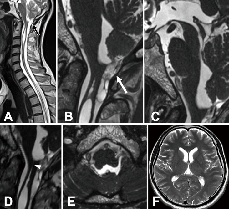
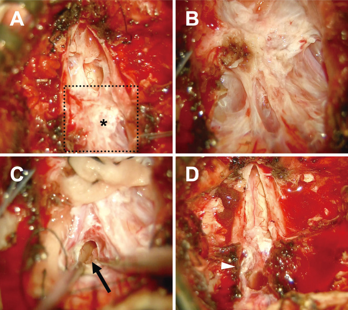
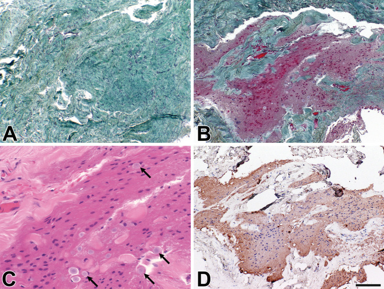
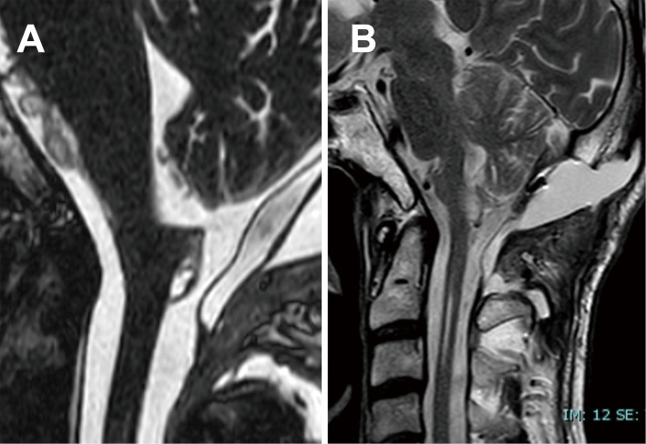
We report a case of syringomyelia in a 55-year-old man with a unique obstruction of Magendie's foramen. Spinal magnetic resonance imaging revealed a large syrinx extending from C1 to C3, with intermittent syringomyelia extending down to Th11. While the obstruction was not clearly evident on T2-weighted imaging, three-dimensional constructive interference in steady-state imaging demonstrated a thickened membranous tissue blocking the cerebrospinal fluid outlet, leading to syrinx formation. Surgical intervention, involving the resection of the thickened membrane to open the foramen of Magendie, resulted in considerable improvement in the syringomyelia and neurological symptoms. Histopathological examination revealed gliomesenchymal tissue, suggesting an embryonic origin of the obstruction. This case highlights the importance of detailed imaging, particularly three-dimensional constructive interference in steady-state sequence, in diagnosing foramen of Magendie obstruction and the potential for successful surgical treatment in selected cases. Histopathological examination is crucial for differentiating gliomesenchymal tissue from adhesive arachnoiditis.

Keywords: 3D-CISS; Magendie's foramen; gliomesenchymal tissue; membranous obstruction; syringomyelia.


Conflict of interest statement

All authors have no conflict of interest.

Figures

Fig. 1
Fig. 1
Preoperative MRI scans. (A) T2-weighted sagittal MRI of the spine shows a large syrinx extending from C1 to C3, with intermittent syringomyelia extending to Th11 and associated spinal cord atrophy. The obstruction of the foramen of Magendie is not clearly visualized (black arrow). Sagittal CISS MRI sequences (B and C) reveal a thickened membrane (white arrow) overlying the foramen of Magendie and inferior enlargement of the fourth ventricle. There is no evidence of aqueductal stenosis or third ventricle dilatation. Reconstructed CISS images (D) show a narrow central canal connecting the syrinx to the fourth ventricle. Axial CISS images (E) show that the foramina of Luschka are normal in size. A T2-weighted axial image of the brain (F) shows that the lateral ventricles are normal in size. CISS: constructive interference in steady-state; MRI: magnetic resonance imaging
Fig. 2
Fig. 2
Intraoperative findings. In all images, the top corresponds to the caudal direction, and the bottom corresponds to the cranial direction. (A) After dural opening, a thickened membranous structure covering the foramen of Magendie (asterisk) was identified. (B) A magnified view of the area enclosed by the dotted line. (C) The membranous structure was dissected, and the fourth ventricle was opened. The choroid plexus within the fourth ventricle is indicated by the black arrow. (D) The remaining thickened membrane was sutured to the right dura mater to prevent obstruction of the cerebrospinal fluid outflow pathway (white arrowhead).
Fig. 3
Fig. 3
Photomicrographs showing histopathological features of the resected membrane. (A) Low-magnification view showing a predominant connective tissue component. (B and D) GFAP-positive glial tissue intermingled within the connective tissue. (C) Higher-magnification view demonstrating astrocytes and numerous corpora amylacea (black arrow). Neurons, inflammatory cells, and neoplastic cells were not identified. Stains used: Elastica–Goldner (A and B), H&E (C), and GFAP staining (D). Scale bars: 225 μm (A), 110 μm (B and D), and 45 μm (C). GFAP: glial fibrillary acidic protein; H&E: hematoxylin and eosin
Fig. 4
Fig. 4
Postoperative MRI scans. (A) Sagittal CISS images show that the foramen of Magendie is widely opened, and the fourth ventricle has decreased in size. (B) T2-weighted sagittal MRI of the spine shows a marked reduction in the size of the syrinx. CISS: constructive interference in steady-state; MRI: magnetic resonance imaging

Similar articles

References

    1. Rifkinson-Mann S, Sachdev VP, Huang YP. Congenital fourth ventricular midline outlet obstruction. Report of two cases. J Neurosurg. 1987;67(4):595-9. doi: 10.3171/jns.1987.67.4.0595 - DOI - PubMed
    1. Huang YC, Chang CN, Chuang HL, et al. Membranous obstruction of the fourth ventricle outlet. A case report. Pediatr Neurosurg. 2001;35(1):43-7. doi: 10.1159/000050385 - DOI - PubMed
    1. Rougier A, Ménégon P. MRI evidence of membranous occlusion of the foramen of Magendie. Acta Neurochir (Wien). 2009;151(6):693-4. doi: 10.1007/s00701-009-0225-5 - DOI - PubMed
    1. Kasapas K, Varthalitis D, Georgakoulias N, et al. Hydrocephalus due to Membranous Obstruction of Magendie's foramen. J Korean Neurosurg Soc. 2015;57(1):68-71. doi: 10.3340/jkns.2015.57.1.68 - DOI - PMC - PubMed
    1. Orakdogen M, Emon ST, Erdogan B, et al. Fourth ventriculostomy in occlusion of the foramen of Magendie associated with Chiari malformation and syringomyelia. NMC Case Rep J. 2015;2(2):72-5. doi: 10.2176/nmccrj.2014-0245 - DOI - PMC - PubMed

Publication types