

Long-lasting effects of lavender exposure on brain resting-state networks in healthy women
- PMID: 40556871
- PMCID: PMC12186306
- DOI: 10.3389/fnins.2025.1555922
Long-lasting effects of lavender exposure on brain resting-state networks in healthy women
Abstract
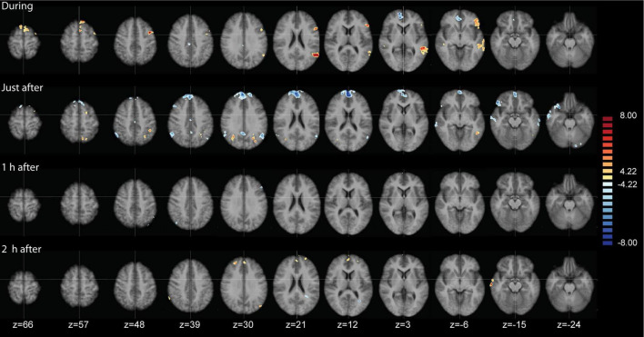
Introduction: Most brain imaging studies on olfaction focus on short-term odorant stimuli, with few examining long-lasting odor exposure or its after-effects. In this study, we utilized resting-state fMRI (rsfMRI) to investigate the effects of prolonged odor exposure to lavender on brain activity and whether these persist post-exposure.
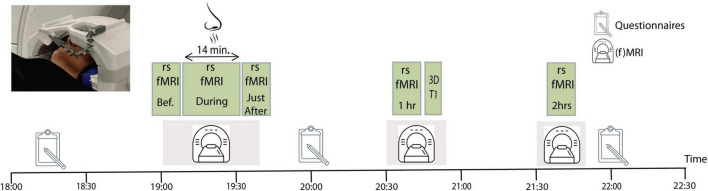
Methods: Fourteen healthy women underwent two fMRI sessions, conducted one week apart, in a randomized order. Both sessions included rsfMRI scans before, during, and up to 2 h after a 14 min exposure to either lavender essential oil or a non-odorant control.
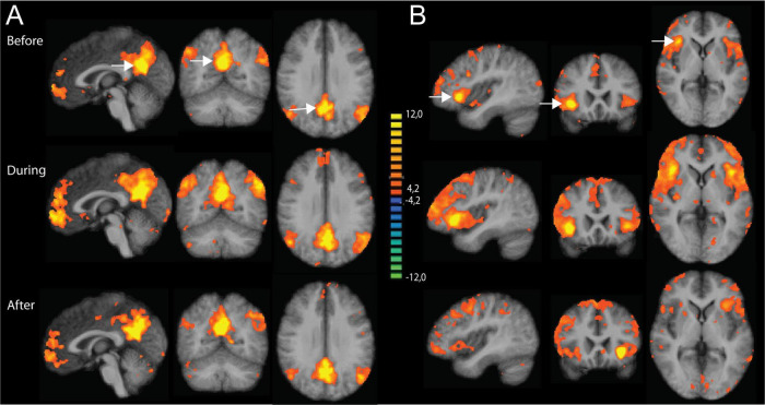
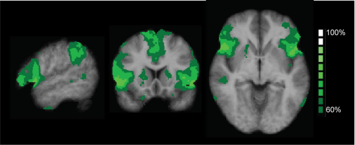
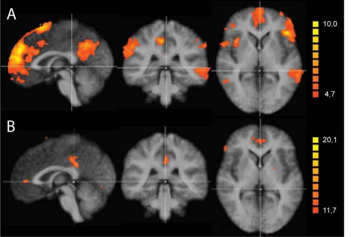
Results: An Independent Component Analysis identified the salience network (SAL) and default mode network (DMN) as the most consistent resting-state networks. A two-factorial ANOVA revealed significant time-varying interaction effects between the SAL and DMN. During odor exposure, functional connectivity (FC) increased within the SAL, and a negative correlation between the SAL and DMN appeared, which intensified immediately after exposure. Two hours post-exposure, the FC between SAL and DMN turned positive.
Discussion: These findings suggest that prolonged odorant exposure to lavender can induce long-lasting brain effects detectable up to 2 h afterwards in women. This proof-of-concept study should be extended to other odorants and to men, and offers new possibilities for exploring the effects of aromatherapy or other odor exposure interventions on brain activity.
Keywords: Independent Component Analysis (ICA); default mode network (DMN); olfactory connectome; resting state functional MRI (rsfMRI); salience network (SAL).
Copyright © 2025 Kupers, Dousteyssier, Delforge, Gonnot, Kantono, Blerot, Pêtre, Dricot and Heinecke.
Conflict of interest statement
OD, JD, AP, and AH were employed by company Brain Impact. RK is member of the the scientific board of Brain Impact. VG and BB were employed by company LMR Naturals By IFF. KK was employed by company IFF. AH was employed by NIRx Medizintechnik Gmbh. The remaining author declares that the research was conducted in the absence of any commercial or financial relationships that could be construed as a potential conflict of interest.
Figures
Similar articles
-
Systematic review and economic analysis of the comparative effectiveness of different inhaled corticosteroids and their usage with long-acting beta2 agonists for the treatment of chronic asthma in adults and children aged 12 years and over.Health Technol Assess. 2008 May;12(19):iii-iv, 1-360. doi: 10.3310/hta12190. Health Technol Assess. 2008. PMID: 18485271
-
Systemic pharmacological treatments for chronic plaque psoriasis: a network meta-analysis.Cochrane Database Syst Rev. 2021 Apr 19;4(4):CD011535. doi: 10.1002/14651858.CD011535.pub4. Cochrane Database Syst Rev. 2021. Update in: Cochrane Database Syst Rev. 2022 May 23;5:CD011535. doi: 10.1002/14651858.CD011535.pub5. PMID: 33871055 Free PMC article. Updated.
-
In mice, discrete odors can selectively promote the neurogenesis of sensory neuron subtypes that they stimulate.Elife. 2025 Jun 18;13:RP96152. doi: 10.7554/eLife.96152. Elife. 2025. PMID: 40531183 Free PMC article.
-
Systemic pharmacological treatments for chronic plaque psoriasis: a network meta-analysis.Cochrane Database Syst Rev. 2017 Dec 22;12(12):CD011535. doi: 10.1002/14651858.CD011535.pub2. Cochrane Database Syst Rev. 2017. Update in: Cochrane Database Syst Rev. 2020 Jan 9;1:CD011535. doi: 10.1002/14651858.CD011535.pub3. PMID: 29271481 Free PMC article. Updated.
-
Interventions to prevent occupational noise-induced hearing loss.Cochrane Database Syst Rev. 2017 Jul 7;7(7):CD006396. doi: 10.1002/14651858.CD006396.pub4. Cochrane Database Syst Rev. 2017. PMID: 28685503 Free PMC article.
References
LinkOut - more resources
Full Text Sources
Miscellaneous