

Alternate quantification approaches for cold-induced vasodilation in human glabrous skin
- PMID: 40556959
- PMCID: PMC12185298
- DOI: 10.3389/fphys.2025.1575764
Alternate quantification approaches for cold-induced vasodilation in human glabrous skin
Abstract
Introduction: Cold-induced vasodilation (CIVD) is a counterintuitive focal increase in glabrous skin blood flow during cold exposure with unclear local and neural mechanisms.
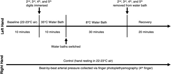
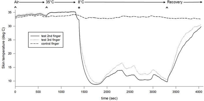
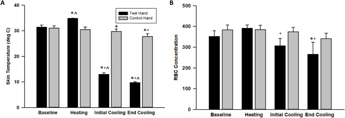
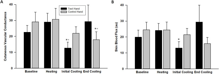
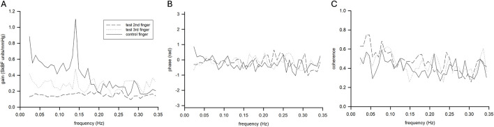
Methods: We tested 12 (8 men, 4 women) healthy subjects' laser-Doppler flux (LDF; just proximal to the nailbed) and arterial blood pressure (ABP) on a beat-by-beat basis. The experimental hand was exposed to warm (10 min 35°C) and then cold (30 min 8°C) water immersion and the contralateral control hand experienced 22°C-23°C air throughout. We analyzed beat-by-beat oscillations in LDF and ABP via a fast-Fourier transform (FFT) and transfer function analysis (TFA) of LDF to ABP.
Results: LDF spectral power was greater in the control finger than immersed fingers in the normalized very low frequency (nVLF) range. There was an interaction in the normalized low frequency (nLF) range where cooling decreased power in immersion sites but increased power in the control site. VLF and LF TFA gains were lower during cooling for immersion but not control sites. Data confirm a significant effect of local vasoconstriction within sympathetic vasoconstriction as identified by changes in VLF and LF, respectively. Comparing CIVD bins (LDF criteria, n = 6) to general cutaneous vasoconstriction bins with no CIVD (n = 6) yielded increased nVLF (P = 0.05) and decreased nLF (P = 0.09) power with CIVD.
Discussion: Thus, the unique analysis of LDF and ABP using the FFT-TFA approach appears to be beneficial in providing insights into CIVD events with a periodic local release of vasoconstriction under varying sympathetic tone.
Keywords: fast-Fourier transform; functional sympatholysis; laser Doppler flowmetry; sympathoexcitation; transfer function analysis.
Copyright © 2025 Stout, Gerow, Clegg, Metzler-Wilson and Wilson.
Conflict of interest statement
The authors declare that the research was conducted in the absence of any commercial or financial relationships that could be construed as a potential conflict of interest. The author(s) declared that they were an editorial board member of Frontiers, at the time of submission. This had no impact on the peer review process and the final decision.
Figures
References
LinkOut - more resources
Full Text Sources