. 2025 Jun 1;18(6):dmm052285.
doi: 10.1242/dmm.052285. Epub 2025 Jun 25.

DWORF expression is reduced in a large animal model of Duchenne muscular dystrophy

Affiliations

DWORF expression is reduced in a large animal model of Duchenne muscular dystrophy

Aaron M Gibson et al. Dis Model Mech. .

Abstract

Duchenne muscular dystrophy (DMD) is a lethal muscle-wasting disease driven by cytosolic calcium overload, which leads to muscle degeneration. Sarco/endoplasmic reticulum calcium ATPase (SERCA), a key regulator of cytosolic calcium levels, exhibits reduced activity in animal models of DMD and human patients. Dwarf open reading frame (DWORF), a positive SERCA regulator, is downregulated in mdx DMD mice, and adeno-associated virus-mediated DWORF overexpression has been shown to ameliorate DMD cardiomyopathy. The canine DMD model provides a crucial bridge for translating findings from mice to humans. To investigate DWORF expression in this model, we developed a canine-specific anti-DWORF antibody, as the existing murine antibody is ineffective. This antibody detected DWORF in human, pig, cat and rabbit muscle, but not in mouse muscle. DWORF was absent in muscle tissues of neonatal normal dogs but highly expressed in those of adult dogs. In DMD-affected dogs aged 8 months or older, DWORF expression was significantly reduced in both cardiac and skeletal muscle. This study establishes a foundation for evaluating DWORF-based gene therapy in the canine DMD model, advancing the potential for clinical translation.

Keywords: DMD; Canine model; DWORF; Duchenne muscular dystrophy; SERCA2a.


Conflict of interest statement

Competing interests D.D. is a member of the scientific advisory board for Sardocor Corp, and an inventor of several issued and filed patents on DMD gene therapy and AAV vectors. R.W.H. is serving on scientific advisory boards for Regeneron Pharmaceuticals-Intellia Therapeutics collaboration, Prevail Therapeutics, Pfizer and Biomarin, and is also receiving funding from Roche. The other authors declare no competing or financial interests.

Figures

Fig. 1.
Fig. 1.
Evaluation of the mouse anti-DWORF antibody. Cross-species evaluation of the mouse anti-DWORF antibody by western blot analysis. The following muscle tissues from adult non-diseased (normal) animals were examined (lane number indicated in parentheses): (1) wild-type mouse heart, (2) DWORF knockout mouse heart, (3) wild-type mouse soleus, (4) DWORF knockout mouse soleus, (5) human soleus, (6) rabbit soleus, (7) pig soleus, (8) cat soleus, (9) dog gastrocnemius and (10) dog heart. KO, knockout; kDa, molecular mass in kilo Dalton; WT, wild type.
Fig. 2.
Fig. 2.
Validation of a canine DWORF antibody. (A) Multi-species alignment of DWORF amino acid sequences performed by Clustal Omega (see Materials and Methods). The epitopes used to generate species-specific antibodies are underlined (mouse, black; dog, red). The symbols below the alignment indicate the degree of conservation of each amino acid: asterisks, fully-conserved residues; colons, strongly similar properties; periods, weakly similar properties. Each amino acid is colored to depict its properties (black, non-polar; green, polar neutral; pink, polar basic; blue, polar acidic). (B,C) Western blot analysis of heart and skeletal muscle from the indicated species using pre-immunized rabbit serum (B) or the purified canine anti-DWORF antibody (C). The following muscle tissues from adult non-diseased (normal) animals were examined (lane number indicated in parentheses): (1) wild-type mouse heart, (2) DWORF knockout mouse heart, (3) wild-type mouse soleus, (4) DWORF knockout mouse soleus, (5) human soleus, (6) rabbit soleus, (7) pig soleus, (8) cat soleus, (9) dog gastrocnemius and (10) dog heart. Arrowhead indicates DWORF.
Fig. 3.
Fig. 3.
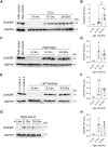
DWORF expression in normal canine muscles from <0.4-month-old, 8- to 13-month-old and 30- to 55-month-old animals. Western blot analysis performed on skeletal muscle and heart tissue from dogs in the following age groups: <0.4 months, 8-13 months and 30-55 months. (A,C,E,G) Representative western blots. (B,D,F,H) Quantification results performed by densitometry with normalization to GAPDH. All data are shown as relative to 8- to 13-month-old samples, where the average of the 8- to 13-month-old group is set to 1. Quantification panels included data from additional blots. (A,B) Extensor carpi ulnaris (ECU) muscle. N=5 for all groups. (C,D) Diaphragm muscle. N=5 for all groups. (E,F) Left ventricle (heart). N=5 for all groups. (G,H) Right atrium (heart). N=1 for <0.4-month-old and N=5 for 8- to 13-month-old and 30- to 55-month-old groups. Data are presented as mean±s.d. *P<0.05 (Wilcoxon rank-sum test). ND, not detected; Rel Exp, relative expression. Skeletal muscle (SkM) control was from the gastrocnemius muscle of a 13-month-old normal dog; diaphragm (Dph) control was from a 13-month-old normal dog; left ventricle (LV) control 1 was from a 12-month-old normal dog; LV control 2 was from a 13-month-old normal dog.
Fig. 4.
Fig. 4.
DWORF expression in age-matched normal and affected canine skeletal muscles. (A-H) Western blot analysis of DWORF expression and quantification in canine extensor carpi ulnaris (ECU) muscle (A-D) and diaphragm (E-H) from normal and affected dogs. The following age groups were examined: <0.4 months (A,E), 8-13 months (B,F) and 30-55 months (C,G). Western blots were quantified by densitometry, normalized to GAPDH and expressed as relative to 8- to 13-month-old normal dogs, where the average of the 8- to 13-month-old normal dogs is set to 1. (D,H) Sample sizes are as follows. ECU: <0.4-month-old, N=5 normal, N=3 DMD; 8 to 13-month-old, N=5 normal, N=4 DMD; 30 to 55-month-old, N=5 normal, N=5 DMD. Diaphragm: <0.4-month-old, N=5 normal, N=4 DMD; 8- to 13-month-old, N=5 normal, N=5 DMD; 30- to 55-month-old, N=5 normal, N=5 DMD. Data are presented as mean±s.d. *P<0.05; **P<0.01 (post-hoc pairwise Wilcoxon rank-sum test with Bonferroni correction). ND, not detected; Rel Exp, relative expression. The positive (+) control sample in A and E is from diaphragm muscle of a 13-month-old normal dog.
Fig. 5.
Fig. 5.
DWORF expression in age-matched normal and affected canine heart tissue. (A-H) Western blot analysis of DWORF expression and quantification in the left ventricle (A-D) and right atrium (E-H) from normal and affected dogs. The following age groups were examined: <0.4 months (A,E), 8-13 months (B,F) and 30-55 months (C,G). Western blots were quantified by densitometry, normalized to GAPDH, and expressed as relative to that in 8- to 13-month-old normal dogs, where the average of the 8- to 13-month-old normal dogs is set to 1 (D,H) Quantification panels included data from additional blots. Sample sizes are as follows. Left ventricle: <0.4-month-old, N=5 normal, N=4 DMD; 8- to 13-month-old, N=7 normal, N=6 DMD; 30- to 55-month-old, N=8 normal, N=12 DMD. Right atrium: <0.4-month-old, N=1 normal, N=4 DMD; 8- to 13-month-old, N=7 normal, N=6 DMD; 30- to 55-month-old, N=8 normal, N=12 DMD. Data are presented as mean±s.d. *P<0.05; **P<0.01; ****P<0.001 (post-hoc pairwise t-test using Tukey's honestly significant difference procedure). ND, not detected; Rel Exp, relative expression. The positive (+) control in A is from the left ventricle of a 12-month-old normal dog, and the+control in panel E is from the left ventricle of a 32-month-old normal dog.

References

    1. Balakrishnan, R., Mareedu, S. and Babu, G. J. (2022). Reducing sarcolipin expression improves muscle metabolism in mdx mice. Am. J. Physiol. Cell Physiol. 322, C260-C274. 10.1152/ajpcell.00125.2021 - DOI - PMC - PubMed
    1. Bers, D. M. (2002). Cardiac excitation-contraction coupling. Nature 415, 198-205. 10.1038/415198a - DOI - PubMed
    1. Bostick, B., Yue, Y., Long, C., Marschalk, N., Fine, D. M., Chen, J. and Duan, D. (2009). Cardiac expression of a mini-dystrophin that normalizes skeletal muscle force only partially restores heart function in aged Mdx mice. Mol. Ther. 17, 253-261. 10.1038/mt.2008.264 - DOI - PMC - PubMed
    1. Bostick, B., Yue, Y. and Duan, D. (2010). Gender influences cardiac function in the mdx model of Duchenne cardiomyopathy. Muscle Nerve 42, 600-603. 10.1002/mus.21763 - DOI - PMC - PubMed
    1. Burr, A. R. and Molkentin, J. D. (2015). Genetic evidence in the mouse solidifies the calcium hypothesis of myofiber death in muscular dystrophy. Cell Death Differ. 22, 1402-1412. 10.1038/cdd.2015.65 - DOI - PMC - PubMed

MeSH terms

Substances

LinkOut - more resources