. 2025 Aug 13;16(8):e0051625.
doi: 10.1128/mbio.00516-25. Epub 2025 Jun 25.

Hybridization capture sequencing for Vibrio spp. and associated virulence factors

Affiliations

Hybridization capture sequencing for Vibrio spp. and associated virulence factors

Kyle D Brumfield et al. mBio. .

Abstract

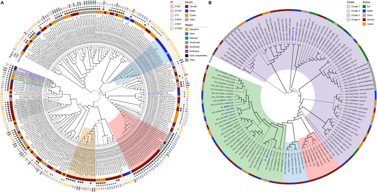
Proliferation of Vibrio spp. in aquatic ecosystems is associated with climate change and, concomitantly, increased incidence of vibriosis. They are autochthonous to aquatic environments globally, but traditional metagenomic methods for detecting and typing pathogenic Vibrio spp. are challenged by their presence in relatively low abundance and ability to persist in a viable but nonculturable state. In the study reported here, hybridization capture sequencing (HCS) was employed to profile low-abundance Vibrio spp. in environmental samples. The HCS panel targeted a family of molecular chaperones (CPN60) specific to 69 Vibrio spp. and 162 Vibrio-specific virulence factors. This approach was evaluated in parallel with traditional whole-community shotgun sequencing in a metagenomic analysis of water and oyster samples collected from the Chesapeake Bay. In addition, Vibrio parahaemolyticus and Vibrio vulnificus strains isolated from the samples were subjected to whole-genome sequencing to determine the genetic characteristics of pathogenic Vibrio spp. circulating in an aquatic environment. HCS, employed to determine the incidence and characterization of specific Vibrio spp., yielded significantly greater metagenomic insight, notably a variety of other Vibrio spp., including detection of Vibrio cholerae, Vibrio fluvialis, and Vibrio aestuarianus, in addition to Vibrio parahaemolyticus and Vibrio vulnificus, and also important virulence factors not detectable using traditional molecular methods. Thus, pathogenic Vibrio spp. in aquatic ecosystems may be far more common than currently understood. It is concluded that environmental surveillance should include HCS, a valuable tool for the detection and characterization of pathogenic agents in aquatic ecosystems, notably vibrios.IMPORTANCEThe increasing prevalence of pathogenic Vibrio spp. in aquatic ecosystems, driven by climate change, is closely linked to a rise in cholera and vibriosis cases, emphasizing the need for improved environmental surveillance. Vibrios are naturally occurring in aquatic environments globally, but traditional metagenomic methods for detecting and typing pathogenic Vibrio spp. are challenged by their presence in relatively low abundance and ability to persist in a viable but nonculturable state. In the study reported here, hybridization capture sequencing was employed to profile low-abundance Vibrio spp. in metagenomic samples, namely water and oysters collected from the Chesapeake Bay. This approach was evaluated in parallel with traditional whole-community shotgun sequencing and whole-genome sequencing of Vibrio parahaemolyticus and Vibrio vulnificus strains isolated from the samples. Results suggest pathogenic Vibrio spp. in aquatic ecosystems may be far more common than currently understood, when multiple methods are considered for environmental surveillance.

Keywords: Vibrio; hybridization capture; metagenomics; microbiome; next generation sequencing; targeted enrichment; virulence; whole-genome sequencing.


Conflict of interest statement

The authors declare no conflict of interest.

Figures

Fig 1
Fig 1
Area of study and PCR characterization. (A) Map of sampling locations in the Chesapeake Bay, Maryland, where water and oyster samples were collected between June and October 2019. (B) Heatmap showing detection (presence/absence) of Vibrio genus- and species-specific biomarkers, including virulence-associated genes, in environmental water and oyster samples as well as pure culture isolates (V. parahaemolyticus and V. vulnificus).
Fig 2
Fig 2
Microbial community composition based on k-mer alignment to whole-genome reference databases. (A) Alpha diversity metrics (observed, Chao1, and inverse Simpson indices) at the species level, comparing water and oyster microbiome profiled by hybridization capture sequencing and shotgun metagenomics. Violin plots are grouped by sample type and colored by sample site. (B) Stacked bar plots showing the relative sequence read abundance (%) of dominant bacterial and archaeal phyla across control, water, and oyster samples. (C) Stacked bar plots showing relative abundance (%) of the top 20 most abundant genera. (D) Heatmap of log-transformed relative abundances, log(RA%), of Vibrio spp. detected across samples and controls, highlighting improved detection sensitivity of hybridization capture sequencing compared to shotgun metagenomics. (E) Heatmap of log-transformed relative abundances, log(RA%), of archaeal, algal, viral, and phage taxa, showing broader taxonomic diversity captured by shotgun metagenomics in water samples. No archaeal, algal, viral, or phage taxa were found in oyster samples; these taxa were combined for each sequencing method (hybridization capture and shotgun metagenomics) to aid visualization. Taxonomic profiling was performed using KMCP with reference databases from GTDB, RefSeq, and GenBank.
Fig 3
Fig 3
CPN60 diversity and phylogenetic analysis of recovered sequences. (A) Taxonomic assignment of CPN60 sequences from metagenomic-assembled contigs across control, water, and oyster samples, using both hybridization capture and shotgun metagenomics. Yellow indicates detection of CPN60 sequences assigned to each taxon; blue indicates not detected. Taxa include Vibrio species, primarily detected by hybridization capture sequencing, as well as non-Vibrio microbial taxa detected by shotgun metagenomics. (B) Neighbor-joining phylogenetic tree of Vibrio spp. CPN60 nucleotide sequences recovered from metagenomics (blue text, starred) and reference sequences (black text) from the chaperonin database. Red dots represent bootstrap support at key nodes. Tree illustrates the phylogenetic placement of environmental sequences among known Vibrio species, confirming species-level assignments and revealing diversity among recovered CPN60 sequences.
Fig 4
Fig 4
Detection and quantification of antimicrobial resistance and virulence factor-associated genes using ShortBRED. (A) Heatmap showing detection and quantification of antimicrobial resistance genes from the CARD, stratified by resistance mechanism. Color scale indicates log-transformed reads per kilobase per million mapped reads, log(RPKMs). (B) Bar plot showing the total count of virulence factor genes detected in each sample, grouped by virulence category based on annotations from the VFDB and probe targets included in the HCS panel. (C) Heatmap showing detection and quantification of virulence factors, including genes from VFDB and custom probe targets, across different sample types and sequencing methods. Virulence genes are grouped by genus (top) and functional category (bottom). Color scale represents log-transformed RPKMs, and VF categories are color-coded in the legend to indicate functional roles.
Fig 5
Fig 5
Phylogenomic relationships of Vibrionaceae isolates and characterization of antimicrobial resistance and virulence factor-associated genes using ShortBRED. (A) Maximum-likelihood phylogenetic tree of Vibrionaceae constructed using single-copy core genes identified by GToTree. Isolates recovered in this study are marked with red stars, and taxonomic groups of interest (V. parahaemolyticus and V. vulnificus) are highlighted in orange and green, respectively. (B) Heatmaps showing detection of VFs in Vibrio spp. isolates. VFs are grouped by functional category according to the VFDB and probe targets included in the HCS panel, with color-coding for functional role. (C) Detection of antimicrobial resistance genes from the CARD database, grouped by resistance mechanism. Yellow indicates the presence, and blue indicates the absence of the target gene or protein family.
Fig 6
Fig 6
Phylogenetic relationships of Vibrio species isolates recovered in this study. Maximum-likelihood phylogenetic tree of (A) Vibrio parahaemolyticus (n = 259 genomes) and (B) Vibrio vulnificus (n = 118 genomes), constructed using single-copy core genes identified by GToTree. Isolates recovered in this study are highlighted in blue and denoted with triangles. Trees were rooted and visualized using iTOL. Clustering illustrates the genomic relatedness of environmental isolates from this study relative to globally distributed reference strains, supporting lineage classification and potential virulence associations.

References

    1. Joseph SW, Colwell RR, Kaper JB. 1982. Vibrio parahaemolyticus and related halophilic vibrios. CRC Crit Rev Microbiol 10:77–124. doi: 10.3109/10408418209113506 - DOI - PubMed
    1. Le Roux F, Blokesch M. 2018. Eco-evolutionary dynamics linked to horizontal gene transfer in vibrios. Annu Rev Microbiol 72:89–110. doi: 10.1146/annurev-micro-090817-062148 - DOI - PubMed
    1. Brumfield K.D, Usmani M, Chen KM, Gangwar M, Jutla AS, Huq A, Colwell RR. 2021. Environmental parameters associated with incidence and transmission of pathogenic Vibrio spp. Environ Microbiol 23:7314–7340. doi: 10.1111/1462-2920.15716 - DOI - PubMed
    1. Brumfield KD, Chen AJ, Gangwar M, Usmani M, Hasan NA, Jutla AS, Huq A, Colwell RR. 2023. Environmental factors influencing occurrence of Vibrio parahaemolyticus and Vibrio vulnificus. Appl Environ Microbiol 89:e0030723. doi: 10.1128/aem.00307-23 - DOI - PMC - PubMed
    1. Huq A, Small EB, West PA, Huq MI, Rahman R, Colwell RR. 1983. Ecological relationships between Vibrio cholerae and planktonic crustacean copepods. Appl Environ Microbiol 45:275–283. doi: 10.1128/aem.45.1.275-283.1983 - DOI - PMC - PubMed

Substances

LinkOut - more resources