. 2025 May 26;32(6):306.
doi: 10.3390/curroncol32060306.

The Value of Thin Layer Cytology in Cancer Patients' Pericardial Effusions

Affiliations

The Value of Thin Layer Cytology in Cancer Patients' Pericardial Effusions

Christos Lafaras et al. Curr Oncol. .

Abstract

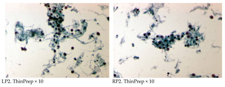
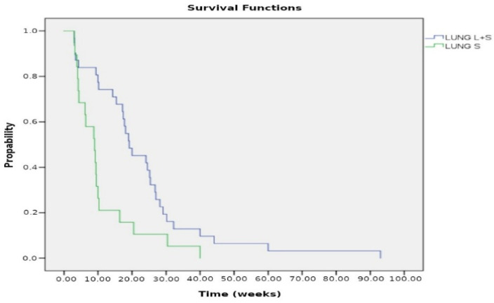
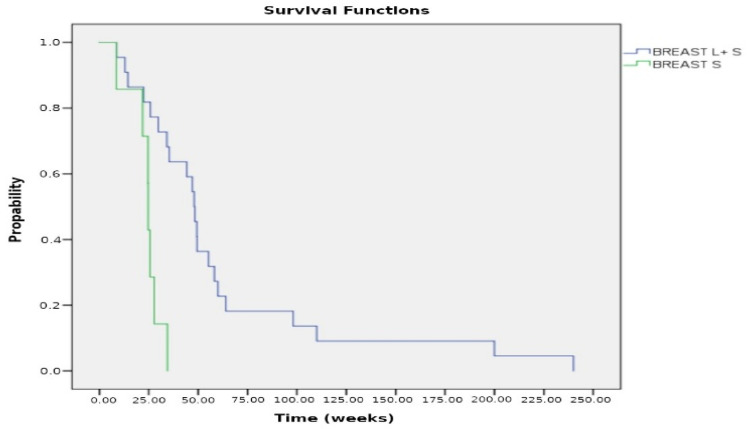
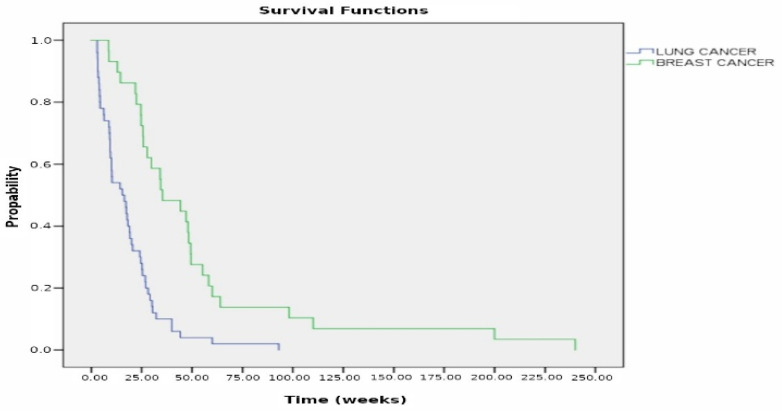
Objective: Early diagnosis and treatment of metastatic pericardial disease are crucial to prevent the life-threatening complication of cardiac tamponade. Thin Layer Cytology (TLC), a widely adopted technique in cytology, has gained significant acceptance for most specimens. Our study aimed to assess the utility of TLC in diagnosing metastatic neoplasms and their origins in pericardial effusions, as well as monitoring response to chemotherapy. Methods: We examined 184 pericardial fluids collected by pericardiocentesis and processed using the ThinPrep liquid-based technique. Various immunocytochemical markers were used to determine the site of metastatic neoplasms. We also evaluated the response to therapy in 53 patients with lung and breast cancer. Results: Out of 184 specimens, 113 pericardial fluids were diagnosed as positive for malignancy, while 71 were negative. Twenty-three cases of unknown primary site were included in the total positive cases. Ninety cases positive for malignancy had a known primary site of origin, including 31 lung carcinomas, 22 breast carcinomas, 10 ovarian carcinomas, 6 T-cell lymphomas, 3 urinary bladder carcinomas, 4 renal carcinomas, 5 adenocarcinomas of the colon, 5 prostate carcinomas, 2 parotid adenocarcinomas, and 2 melanomas. Regarding the 53 cases with chemotherapy treatment, the cytologic examination of pericardial fluid showed a remarkable reduction in neoplastic burden after the third dose of cisplatin or thiotepa instilled into the pericardial cavity. ThinPrep provided excellent preservation of cytomorphological features, high cellularity per slide, and a clear background. This comprehensive analysis provides crucial information about the types and distribution of cancerous cells present in the samples. Conclusions: Thin Layer Cytology (TLC) is a valuable diagnostic tool for detecting metastatic pericardial malignancy. It allows the examination of exfoliated cells from the pericardial fluid, providing crucial information for diagnosis, management, and monitoring the acute responsiveness to intrapericardial chemotherapy. Immunocytochemistry (IHC) can identify specific markers for various types of cancer, enabling a more accurate diagnosis and guiding further treatment decisions.

Keywords: Thin Layer Cytology; ThinPrep; cardiac tamponade; immunocytochemistry; liquid based cytology; metastatic pericardial effusions.


Conflict of interest statement

The authors declare no conflicts of interest.

Figures

Figure 1
Figure 1
Cytomorphological image indicative of metastatic adenocarcinoma (ACA) of lung in pericardial effusion. High cellularity with numerous atypical cells, neoplastic cells as clusters or singly dispersed, nuclear pleomorphism. (A). Papstain (reveals nuclear details—membrane irregularities, chromatin pattern, and intranuclear invaginations, ThinPrep ×400. (B). NapsinA (Immunohistochemistry for Nap-A, a functional aspartic proteinase, as a marker for primary lung ACA. ThinPrep ×400.
Figure 2
Figure 2
Cytomorphological image of metastatic ductal adenocarcinoma (invasive breast carcinoma, NST) of the breast in pericardial effusion. (A). Papstain, ThinPrep ×400. (B). GATA3 (nuclear staining) positive, positive in ductal adenocarcinoma, ThinPrep ×400.
Figure 3
Figure 3
Cytomorphological image of metastatic adenocarcinoma of the colon in pericardial effusion. (A). Papstain, ThinPrep ×400. (B). CDX2 positive in adenocarcinoma of colon, ThinPrep ×400.
Figure 4
Figure 4
Cytological examination of pericardial fluid before (left panel, LP1, LP2) and after the 3rd dose of intrapericardial administration of cisplatin (right panel, RP1, RP2) in lung cancer patients with malignant cardiac tamponade. A reduced neoplastic burden is depicted by decreased and smaller sample-sized groups of malignant cells in these smears.
Figure 4
Figure 4
Cytological examination of pericardial fluid before (left panel, LP1, LP2) and after the 3rd dose of intrapericardial administration of cisplatin (right panel, RP1, RP2) in lung cancer patients with malignant cardiac tamponade. A reduced neoplastic burden is depicted by decreased and smaller sample-sized groups of malignant cells in these smears.
Figure 5
Figure 5
Kaplan–Meier curves for patients with lung cancer comparing combined local and systemic chemotherapy (L + S) versus systemic chemotherapy alone (S). The median survival was 19.2 weeks for the L + S treatment compared to 9 weeks for the S treatment; p < 0.005.
Figure 6
Figure 6
Kaplan–Meier curves for patients with breast cancer who received combined chemotherapy: Local plus systemic (L + S) vs. systemic chemotherapy (S) alone. The median survival was 48 weeks for the L + S treatment compared to 24.7 weeks for the S treatment; p = 0.001.
Figure 7
Figure 7
Kaplan–Meier curves of observation in patients with breast cancer vs. lung cancer. The median survival was 15.4 weeks for lung cancer vs. 35.4 weeks for breast cancer; p < 0.00005.

Similar articles

References

    1. Mori S., Bertamino M., Guerisoli L., Stratoti S., Canale C., Spallarossa P., Porto I., Ameri P. Pericardial effusion in oncological patients: Current knowledge and principles of management. Cardiooncology. 2024;10:8. doi: 10.1186/s40959-024-00207-3. - DOI - PMC - PubMed
    1. Chahine J., Shekhar S., Mahalwar G., Imazio M., Collier P., Klein A. Pericardial Involvement in Cancer. Am. J. Cardiol. 2021;145:151–159. doi: 10.1016/j.amjcard.2020.12.092. - DOI - PubMed
    1. Moores D.W., Dziuban S.W. Pericardial drainage procedures. Chest Surg. Clin. N. Am. 1995;5:359–373. doi: 10.1016/S1052-3359(25)00697-0. - DOI - PubMed
    1. Marcy P.Y., Bondiau P.Y., Brunner P. Percutaneous treatment in patients presenting with malignant cardiac tamponade. Eur. Radiol. 2005;15:2000–2009. doi: 10.1007/s00330-004-2611-y. - DOI - PubMed
    1. Marcy P.Y., Magné N. Imaging of percutaneous pericardioperitoneal shunt in patient with malignant cardiac tamponade. JBR-BTR. 2004;87:186–189. - PubMed

LinkOut - more resources