

Examination and Relationship of Posterior Superior Alveolar Artery and Canalis Sinuosus Using Cone Beam CT
- PMID: 40558321
- PMCID: PMC12191117
- DOI: 10.3390/biomimetics10060352
Examination and Relationship of Posterior Superior Alveolar Artery and Canalis Sinuosus Using Cone Beam CT
Abstract
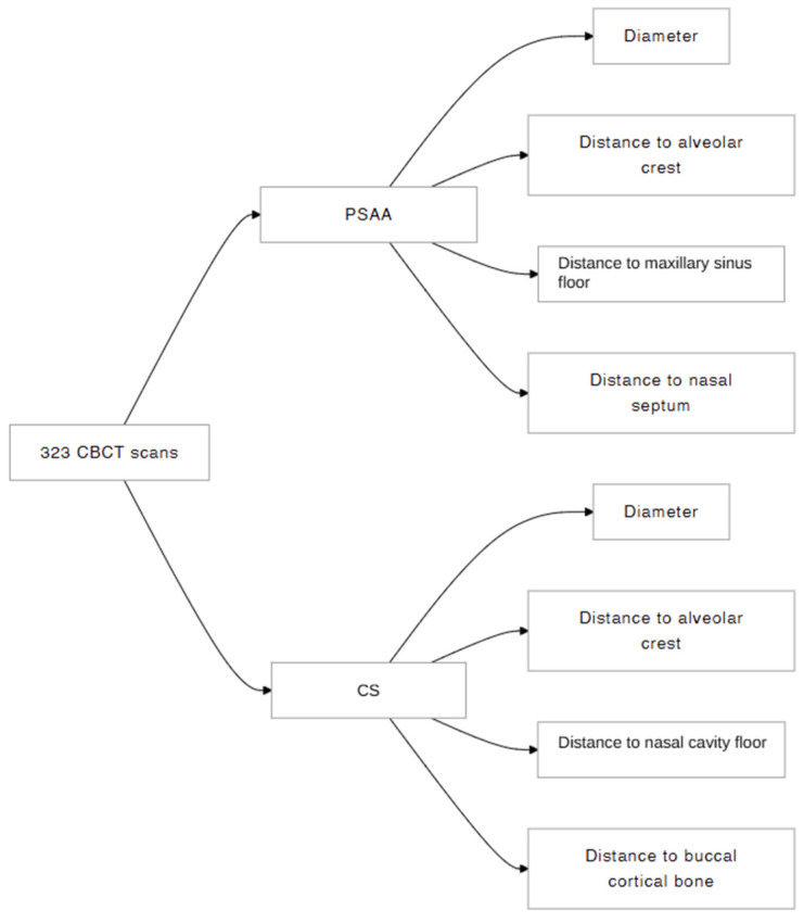
In this study, we investigated the anatomical location, dimensions, and relationships of the posterior superior alveolar artery (PSAA) and canalis sinuosus (CS) within the maxilla, aiming to enhance the safety and efficacy of surgical procedures. A retrospective analysis was performed on 323 individual cone beam computed tomography scans. The diameter of the PSAA and CS, the distance of the PSAA from the sinus floor, the distance of the PSAA and CS from the alveolar crest, the distance of the PSAA and CS from the nasal septum, and the distance from CS to the nasal cavity floor were measured. The distance between PSAA and the sinus floor showed no significant difference between the right and left sides nor between genders (p < 0.05). The distance between the alveolar crest of PSAA and the distance between PSAA and to nasal septum was significantly higher on the left than on the right side (p < 0.05). According to gender, female subjects exhibited a lower distance between PSAA and the nasal septum than males (p < 0.05). Variations in PSAA and CS anatomy highlight the need for individualized preoperative CBCT assessment to reduce complications like bleeding during maxillary surgeries, enhancing surgical planning and safety in dental and maxillofacial procedures.
Keywords: anatomy; canalis sinuosus; cone beam computed tomography; maxilla; posterior superior alveolar artery.
Conflict of interest statement
The authors declare no conflicts of interest.
Figures


Similar articles
-
Prevalence of canalis sinuosus and accessory canals of canalis sinuosus on cone beam computed tomography: a systematic review and meta-analysis.Int J Oral Maxillofac Surg. 2023 Jan;52(1):118-131. doi: 10.1016/j.ijom.2022.06.011. Epub 2022 Jul 13. Int J Oral Maxillofac Surg. 2023. PMID: 35840447
-
Detection of the posterior superior alveolar artery in the lateral sinus wall using computed tomography/cone beam computed tomography: a prevalence meta-analysis study and systematic review.Int J Oral Maxillofac Surg. 2015 Nov;44(11):1405-10. doi: 10.1016/j.ijom.2015.07.001. Epub 2015 Jul 26. Int J Oral Maxillofac Surg. 2015. PMID: 26215383
-
[The influence of the relationship between the root apex of the posterior teeth and the maxillary sinus floor on the thickness of the maxillary sinus mucosa].Shanghai Kou Qiang Yi Xue. 2025 Apr;34(2):196-201. Shanghai Kou Qiang Yi Xue. 2025. PMID: 40550774 Chinese.
-
Frequency, location, and diameter of the anastomosis between the posterior superior alveolar artery and the infraorbital artery in imaging studies: systematic review and meta-analysis.Surg Radiol Anat. 2023 Apr;45(4):431-443. doi: 10.1007/s00276-023-03091-1. Epub 2023 Feb 9. Surg Radiol Anat. 2023. PMID: 36754890
-
Comparison of ultrasonography and cone-beam computed tomography for quantitative assessment of mental foramen and alveolar crest.BMC Oral Health. 2025 Jul 2;25(1):1020. doi: 10.1186/s12903-025-06396-2. BMC Oral Health. 2025. PMID: 40604677 Free PMC article.
References
-
- Kasahara N., Morita W., Tanaka R., Hayashi T., Kenmotsu S., Ohshima H. The Relationships of the Maxillary Sinus With the Superior Alveolar Nerves and Vessels as Demonstrated by Cone-Beam CT Combined With μ-CT and Histological Analyses. Anat. Rec. 2016;299:669–678. doi: 10.1002/ar.23327. - DOI - PubMed
LinkOut - more resources
Full Text Sources
