Review
. 2025 Jun 4;15(6):359.
doi: 10.3390/bios15060359.

Surface Plasmon Resonance Aptasensors: Emerging Design and Deployment Landscape

Affiliations
Review

Surface Plasmon Resonance Aptasensors: Emerging Design and Deployment Landscape

Fahd Khalid-Salako et al. Biosensors (Basel). .

Abstract

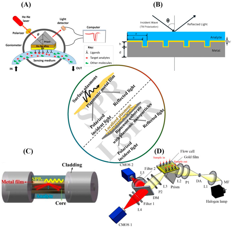
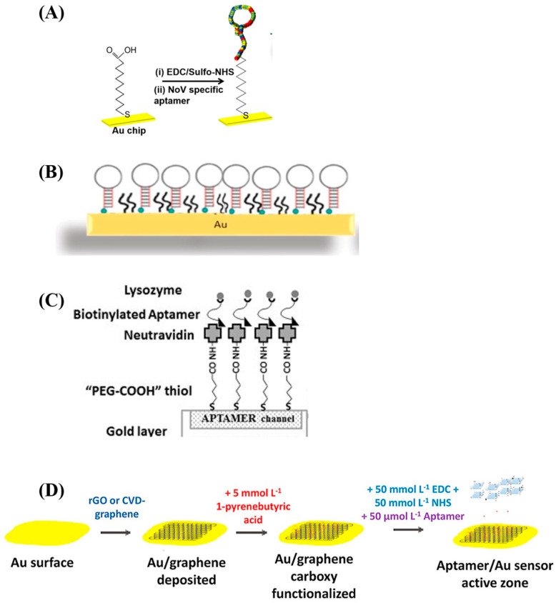
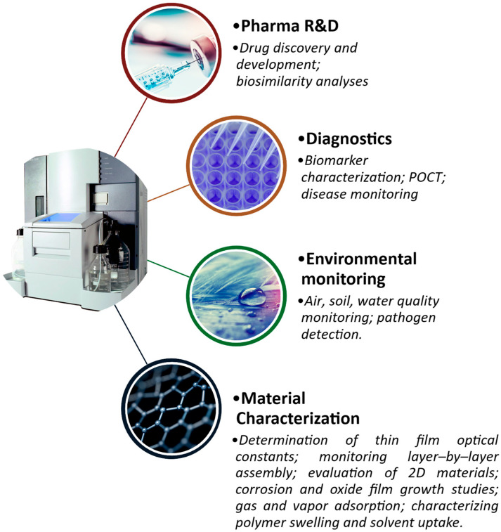
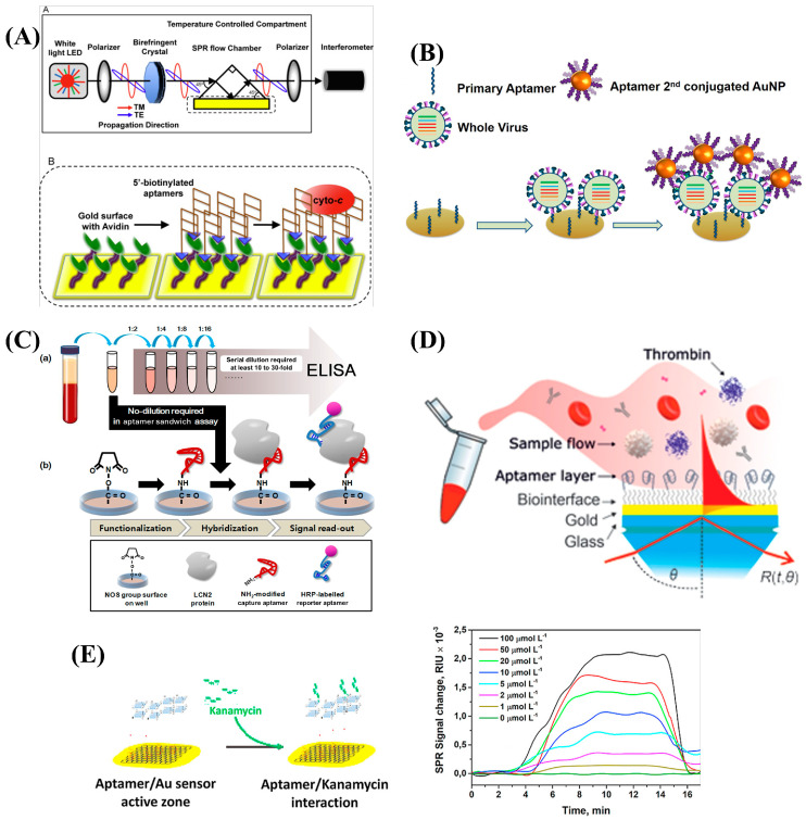
SPR biosensors operate on the principle of evanescent wave propagation at metal-dielectric interfaces in total internal reflection conditions, with consequent photonic energy attenuation. This plasmonic excitation occurs in specific conditions of incident light wavelength, angle, and the dielectric refractive index. This principle has been the basis for SPR-based biosensor setups wherein mass/concentration-induced changes in the refractive indices of dielectric media reflect as plasmonic resonance condition changes quantitatively reported as arbitrary response units. SPR biosensors operating on this conceptual framework have been designed to study biomolecular interactions with real-time readout and in label-free setups, providing key kinetic characterization that has been valuable in various applications. SPR biosensors often feature antibodies as target affinity probes. Notably, the operational challenges encountered with antibodies have led to the development of aptamers-oligonucleotide biomolecules rationally designed to adopt tertiary structures, enabling high affinity and specific binding to a wide range of targets. Aptamers have been extensively adopted in SPR biosensor setups with promising clinical and industrial prospects. In this paper, we explore the growing literature on SPR setups featuring aptamers, specifically providing expert commentary on the current state and future implications of these SPR aptasensors for drug discovery as well as disease diagnosis and monitoring.

Keywords: SELEX; aptamers; biosensors; drug discovery; ligands; surface plasmon resonance.


Conflict of interest statement

The authors declare no conflicts of interest.

Figures

Figure 1
Figure 1
An illustration of the major SPR biosensor configurations. The inset diagram demonstrates the general principle of SPR and LSPR, juxtaposing film-confined evanescent waves in SPR with localized plasmons in LSPR upon polarized light irradiation. (A) A schematic of the conventional Kretschmann SPR configuration. Reproduced with permission from ref. [34]. Copyright 2025 Elsevier B.V. (B) A typical diffraction grating-based SPR setup. p, grating period; b, grating bar width; h, grating thickness; h’, plasmonic metal coating thickness; d, substrate thickness; θ, incident angle (slight modifications made for editorial and formatting purposes) [19]. (C) A diagrammatic sketch of a transmissive fiber optic SPR sensor. SPP, Surface plasmon polaritons. Reproduced with permission from ref. [20]. Copyright 2022, Wiley-VCH GmbH. (D) An SPR imaging (SPRi) system based on the Kretschmann configuration. L1–L5, lenses; DA, diaphragm aperture; MF, multimode fiber; DM, dichroic mirror; P1 and P2, polarizers [35].
Figure 3
Figure 3
Illustrations of various SPR aptasensor surface preparation strategies. (A) The immobilization of a Norovirus capsid aptamer molecule on an SPR chip by amine coupling. The sensor chip is modified with mercaptoundecanoic acid (MUA) which is anchored to the gold film by thiol–gold (S—Au) covalent bonding. The carboxylic acid group of the MUA molecule is activated by EDC/NHS, after which the 5’amine aptamer is reacted with the activated carboxylate. Reproduced with permission from ref. [65]. Copyright 2018, Elsevier B.V. (B) A whole-cell Staphylococcus aureus aptamer immobilized on an SPR chip by thiol coupling. Adapted with permission from ref. [23]. Copyright 2020 American Chemical Society. (C) A lysozyme aptamer immobilized by high-affinity capture. Thiolated PEG-COOH is self-assembled on the chip surface, after which neutravidin is coated on the PEGylated surface by amine coupling. The biotinylated aptamers are then captured through high-affinity neutravidin–biotin capture. Reused with permission from ref. [66]. Copyright 2014 Elsevier B.V. (D) A multi-step procedure for the preparation of an SPR aptasensor surface. Graphene is deposited on the gold surface by chemical vapor deposition, after which PBA is π-stacked and its carboxylic groups are activated for the amine coupling of the NH2-functionalized aptamers. Reproduced with permission from ref. [60]. Copyright 2021 Elsevier B.V.
Figure 2
Figure 2
SPR biosensors have found widespread use in pharmaceutical research and development, clinical diagnostics, environmental monitoring, and the characterization of thin films’ structural and optical properties.
Figure 4
Figure 4
Some examples of SPR aptasensor applications in drug screening and infectious pathogen, disease biomarker, and small-molecule detection. (A) An SPR aptasensor for anti-cancer drug screening. Reproduced with permission from ref. [91]. Copyright 2014, Elsevier B.V. (B) A dual-aptamer SPR biosensor of whole avian flu virus with gold nanoparticle conjugation for signal amplification. Reproduced with permission from ref. [67]. Copyright 2016 Elsevier B.V. (C) A sandwich detection-based SPR aptasensor designed for sandwich immunoassay for Hepatocellular Carcinoma diagnosis [92]. (D) An SPR aptasensor for thrombin detection in human blood. Reproduced with permission from ref. [80]. Copyright 2020, Elsevier B.V. (E) Kanamycin residues detection in food by an SPR aptasensor. Reproduced with permission from ref. [60]. Copyright 2021 Elsevier B.V.

Similar articles

References

    1. Stuart D.D., Van Zant W., Valiulis S., Malinick A.S., Hanson V., Cheng Q. Trends in Surface Plasmon Resonance Biosensing: Materials, Methods, and Machine Learning. Anal. Bioanal. Chem. 2024;416:5221–5232. doi: 10.1007/s00216-024-05367-w. - DOI - PubMed
    1. Wang Y., Liu X., Wu L., Ding L., Effah C.Y., Wu Y., Xiong Y., He L. Construction and Bioapplications of Aptamer-Based Dual Recognition Strategy. Biosens. Bioelectron. 2022;195:113661. doi: 10.1016/j.bios.2021.113661. - DOI - PubMed
    1. Ellington A.D., Szostak J.W. In Vitro Selection of RNA Molecules That Bind Specific Ligands. Nature. 1990;346:818–822. doi: 10.1038/346818a0. - DOI - PubMed
    1. Yüce M., Ullah N., Budak H. Trends in Aptamer Selection Methods and Applications. Analyst. 2015;140:5379–5399. doi: 10.1039/C5AN00954E. - DOI - PubMed
    1. Kulabhusan P.K., Hussain B., Yüce M. Current Perspectives on Aptamers as Diagnostic Tools and Therapeutic Agents. Pharmaceutics. 2020;12:646. doi: 10.3390/pharmaceutics12070646. - DOI - PMC - PubMed

Substances

LinkOut - more resources