

The Calcium Signalling Profile of the Inner Blood-Retinal Barrier in Diabetic Retinopathy
- PMID: 40558483
- PMCID: PMC12191068
- DOI: 10.3390/cells14120856
The Calcium Signalling Profile of the Inner Blood-Retinal Barrier in Diabetic Retinopathy
Abstract
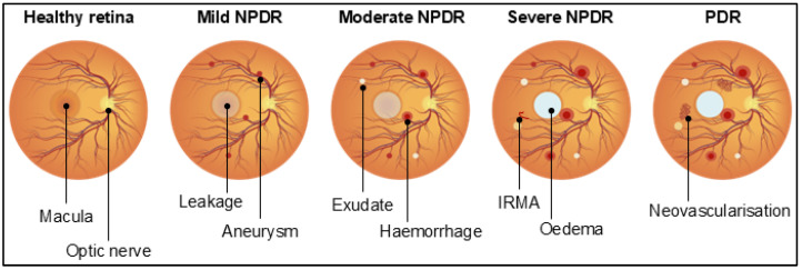
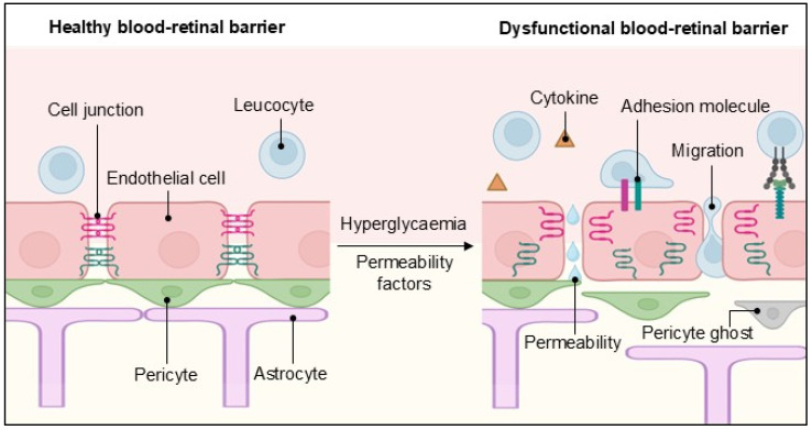
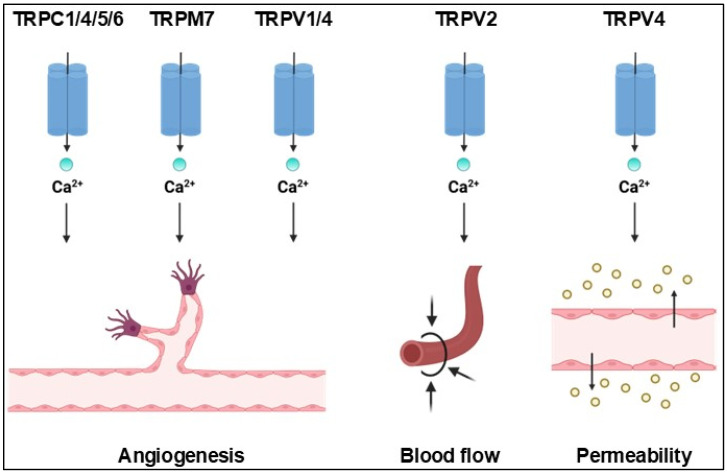
Diabetic retinopathy is a sight-threatening complication of diabetes mellitus, affecting millions of people worldwide. From a vascular perspective, diabetic retinopathy compromises the structure and function of the blood-retinal barrier, leading to aberrant angiogenesis and vascular leakage, with consequent loss of vision. This review will delve into the vascular abnormalities caused by diabetic retinopathy in the inner blood-retinal barrier, focusing primarily on retinal endothelial cells. It will then discuss how calcium signalling regulates inner blood-retina barrier function and dysfunction, how calcium channels contribute to the development of diabetic retinopathy, and how studying the components of the calcium toolkit may identify new therapeutic targets.
Keywords: blood–retinal barrier; calcium signalling; diabetic retinopathy; endothelial dysfunction.
Conflict of interest statement
The authors declare no conflict of interest.
Figures
Similar articles
-
Exploring the role of Müller cells-derived exosomes in diabetic retinopathy.Microvasc Res. 2024 Jul;154:104695. doi: 10.1016/j.mvr.2024.104695. Epub 2024 May 8. Microvasc Res. 2024. PMID: 38723843 Free PMC article.
-
Dectin-1 Drives Diabetic Retinopathy via Inducing Microglia-Mediated Inflammation and Blood-Retinal Barrier Breakdown.Dev Neurobiol. 2025 Oct;85(4):e22997. doi: 10.1002/dneu.22997. Dev Neurobiol. 2025. PMID: 40790981
-
Fenofibrate for diabetic retinopathy.Cochrane Database Syst Rev. 2023 Jun 13;6(6):CD013318. doi: 10.1002/14651858.CD013318.pub2. Cochrane Database Syst Rev. 2023. PMID: 37310870 Free PMC article.
-
Microglia Promote Endothelial Cell Activation Through NSUN2-Mediated SQLE m5C Modification in Diabetic Retinopathy.FASEB J. 2025 Jun 30;39(12):e70742. doi: 10.1096/fj.202500302RR. FASEB J. 2025. PMID: 40536094
-
Optical coherence tomography (OCT) for detection of macular oedema in patients with diabetic retinopathy.Cochrane Database Syst Rev. 2011 Jul 6;(7):CD008081. doi: 10.1002/14651858.CD008081.pub2. Cochrane Database Syst Rev. 2011. Update in: Cochrane Database Syst Rev. 2015 Jan 07;1:CD008081. doi: 10.1002/14651858.CD008081.pub3. PMID: 21735421 Updated.
References
-
- Antar S.A., Ashour N.A., Sharaky M., Khattab M., Ashour N.A., Zaid R.T., Roh E.J., Elkamhawy A., Al-Karmalawy A.A. Diabetes mellitus: Classification, mediators, and complications; A gate to identify potential targets for the development of new effective treatments. Biomed. Pharmacother. 2023;168:115734. doi: 10.1016/j.biopha.2023.115734. - DOI - PubMed
Publication types
MeSH terms
Substances
LinkOut - more resources
Full Text Sources
Medical
