. 2025 Jun 9;14(12):868.
doi: 10.3390/cells14120868.

Regulation of Myogenesis by MechanomiR-200c/FoxO3 Axis

Affiliations

Regulation of Myogenesis by MechanomiR-200c/FoxO3 Axis

Junaith S Mohamed et al. Cells. .

Abstract

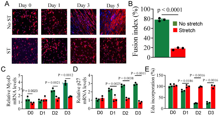
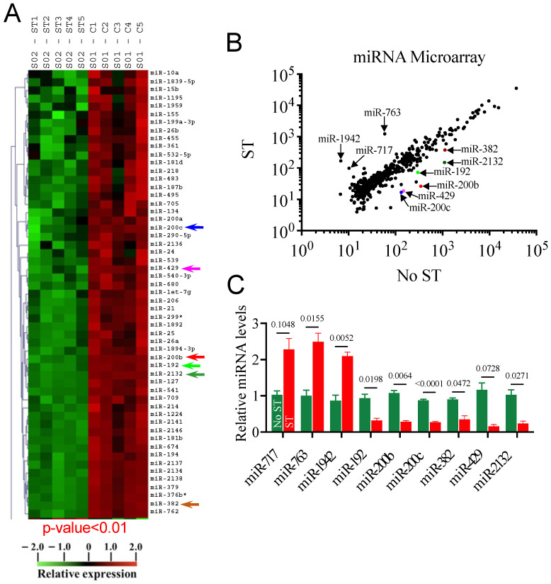
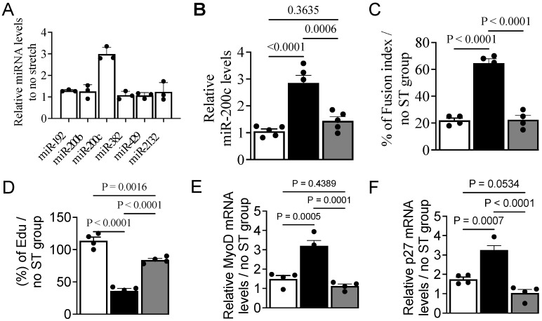
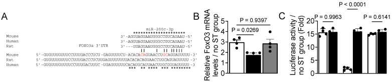
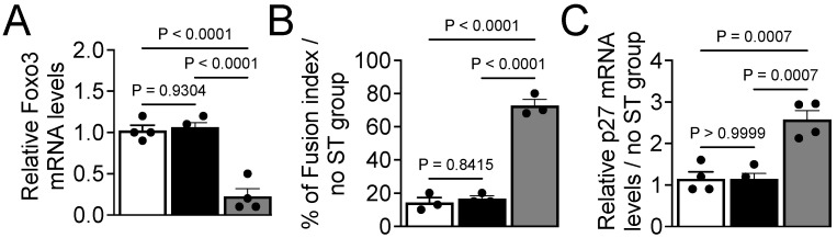
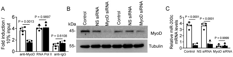
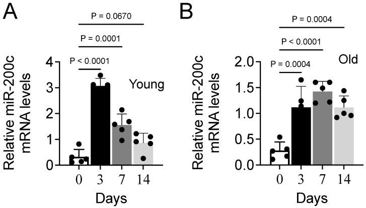
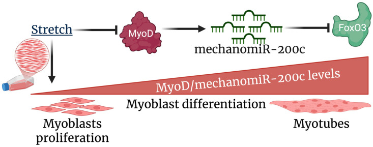
Cyclic mechanical stretch has been shown to inhibit myoblast differentiation while promoting proliferation. However, the underlying molecular mechanisms are not well understood. Here, we report that mechanical stretch inhibits the differentiation of mouse primary myoblasts by promoting the cell cycle program and by inhibiting the expression of the myogenic regulator MyoD. Stretch alters the miRNA expression profile as evidenced by miRNA microarray analysis. We identified miR-200c as one of the highly downregulated mechanosensitive miRNAs (mechanomiRs) whose expression level was increased during differentiation. This suggests that mechanomiRs-200c is a myogenic miRNA. Overexpression of mechanomiR-200c revoked the effect of stretch on myoblast differentiation, and the introduction of the mechanomiR-200c antagomir restored the stretch effect. This suggests that stretch blocks differentiation, in part, through mechanomiR-200c. The gene encoding the transcription factor FoxO3 is a known direct target of mechanomiR-200c. Interestingly, MyoD binds to the mechanomiR-200c promoter in differentiating myoblasts, whereas stretch appears to reverse such binding. Our data further demonstrate that the levels of mechanomiR-200c are robustly elevated during the early stage of the muscle repair process in young mice, but not in the injured muscle of aged mice. Overall, we identified a novel pathway, MyoD/mechanomiR-200c/FoxO3a, and the potential mechanism by which stretch inhibits myoblast differentiation.

Keywords: MyoD; differentiation; mechanical stretch; microRNAs; myoblasts.


Conflict of interest statement

The authors declare no conflicts of interest.

Figures

Figure 1
Figure 1
The effect of stretch on myoblast differentiation. Myoblasts cultured in GM were transferred to DM ~70–80% confluence (considered as day 1) followed by 1 h cyclic stretching for 3 or 5 days. (A) The cells were stained with desmin antibody and DAPI for the indicated periods and imaged at 10X under a fluorescent microscope. (B) The fusion index was calculated on day 5. (C,D) Total RNA was isolated from the differentiating myoblasts at the indicated periods and used in the RT-qPCR to calculate MyoD (C) and p27 (D) mRNA expression levels. Tubulin was used as a normalizer in the qPCR. (E) Cell proliferation was measured by % incorporation of fluorescently labeled thymidine analog (EdU) at the indicated periods. Each bar represents the mean values ± SEM (n = 3/group).
Figure 2
Figure 2
Genome-wide mechanosensitive miRNA expression profile. Myoblasts were subjected to no stretch or 1 h stretch immediately after being transferred to DM. After 12 h, total RNA was isolated and used in miRNA microarray analyses to identify differentially regulated mechanomiRs. (A) A heat map of miRNA microarray analysis showing differential expression of mechanomiRs in the presence (ST) or absence (C) of stretch. The arrows indicate the highly downregulated mechanomiRs. (B) A scatter plot showing log10-transformed signal intensities for Cy3-labeled probes in unstretched myoblasts and Cy5-labeled probes in stretched myoblasts. Each dot represents one miRNA probe. Color dots indicate the highly downregulated mechanomiRs. (C) A portion of the RNA used in the microarray was utilized in RT-qPCR to confirm the expression levels of the selected mechanomiRs that were differentially regulated by >1.5-fold in the microarray. Each bar represents the mean ± SEM (n = 3/group).
Figure 3
Figure 3
MechanomiR-200c promotes myoblast differentiation. (A) Total RNA was isolated from myoblasts 24 h after the switch to DM and used in RT-qPCR to determine the expression levels of mechanomiRs (n = 3). (B) Myoblasts growing in GM were transfected with the pEGP-miR-200c-overexpressing vector (black bar) with or without the anti-miR-200c antagomir (co-transfection; gray bar) or pEGP (empty) vector (white bar) for 24 h. Total RNA was isolated and used in RT-qPCR to determine miR-200c expression. (CF) The transfected myoblasts were cultured in DM to induce differentiation for five days with or without stretch. Immunocytochemistry was performed on day five to calculate the fusion index (C). Fluorescently labeled EdU was added in the above transfected myoblasts immediately after switching to DM to determine the rate of cell proliferation on day 3 by gauging the % incorporation of EdU (D). Total RNA was isolated from the myoblasts and used in qPCR to determine the relative of levels MyoD (E) and p27 (F) mRNA expression. Tubulin was used as a normalizer in the qPCR. Each bar represents the fold difference of the mean ± SEM (n = 4/group) calculated according to the no-stretch group, except ‘B’.
Figure 4
Figure 4
FoxO3a mRNA is a target gene of mechanomiR-200c. (A) Sequence alignment of the putative miR-200c targeting site in the 3′-UTR of FoxO3 shows high levels of complementarity. The star (*) indicates conserved nucleotides, while nucleotides labeled in red indicate the mutated region of the 3′UTR of FoxO3 mRNA. (B) Myoblasts growing in GM were transfected with the pEGP-miR-200c-overexpressing vector (black bar) with or without the anti-miR-200c antagomir (gray bar) or pEGP (white bar) vector for 24 h. Total RNA was isolated from myoblasts 48 h after switching from GM to DM with or without the stretch stimulus and was used in RT-qPCR to determine the expression levels of FoxO3a mRNA levels. (C) Myoblasts in GM were transfected with plasmids that contain the firefly luciferase coding region cloned downstream of a wildtype (white bar) or mutant (black bar) FoxO3a 3′-UTR in the presence of the pEGP empty vector, a pEGP-miR-200c-overexpressing vector. After 48 h of myogenic induction (switched to DM), myoblasts were collected, lysed, and used for measurement of firefly and Renilla luciferase activities in a luminometer. Firefly luciferase activities were normalized to that of Renilla luciferase and plotted. Each bar represents the fold difference of the mean ± SEM (n = 4/group) calculated according to controls (no-stretch group), except ‘C’.
Figure 5
Figure 5
FoxO3 is necessary for the inhibitory effect of stretch on myoblast differentiation. (A) Myoblasts growing in GM were transfected with FoxO3 siRNA (gray bar), non-specific siRNA (black bar), or no transfection (white bar) for 24 h. Total RNA was isolated from myoblasts and used in RT-qPCR to determine the expression levels of FoxO3a mRNA levels. (B) The above transfected myoblasts were transferred from GM to DM to induce differentiation for five days with or without stretch. Immunocytochemistry was performed on day five to calculate the fusion index. (C) Total RNA was isolated from the above transfected myoblasts and used in the immunoblot assay to determine the relative levels of p27 mRNA expression on day five. Tubulin mRNA expression levels were used as a normalizer in the qPCR. Each bar represents the fold difference of the mean ± SEM (n = 4/group) calculated according to controls (no-stretch group), except ‘A’.
Figure 6
Figure 6
MyoD binds to the miR-200c promoter. (A) Myoblasts were cultured for 24 h in DM, subjected to either stretch (black bar) or no stretch (white bar), and chromatin was isolated and precipitated with anti-MyoD, anti-RNA polymerase II (positive control), or non-specific anti-IgG (negative control) antibodies. Occupancy on the miR-200c promoter by MyoD and on the GAPDH promoter by RNA polymerase II was determined using qPCR with primers specific for the miR-200c or GAPDH promoter. (B) Myoblasts growing in GM were transfected with MyoD siRNA, non-specific siRNA, or no transfection for 24 h. Cells were harvested and lysed, and MyoD protein levels were determined by an immunoblot assay. The gel images shown are representative of three independent experiments. (C) The above transfected myoblasts were cultured in DM for three days with (black bars) or without (white bars) stretch. Total cellular RNA was isolated and the levels of miR-200c were determined by RT-qPCR. Tubulin mRNA expression levels were used as a normalizer in the qPCR or loading control in the immunoblot assay. Each bar represents the fold difference of the mean ± SEM (n = 4/group) calculated according to controls (no-stretch group).
Figure 7
Figure 7
The expression of miR-200c during the muscle repair process in young and aged mice. (A,B) The tibialis anterior muscle of young and aged mice was injected with cardiotoxin to induce injury. At various time points after injury (0, 3, 7, or 14 days), the mice were euthanized, and their tibialis anterior muscles were collected. Total cellular RNA was isolated from each muscle and the levels of miR-200c in young (A) or aged (B) mice were determined using RT-qPCR. Each bar indicates the mean values ± SEM (n = 4 for young and 5 for aged mice).
Figure 8
Figure 8
The proposed model of myogenesis following stretch.

Similar articles

References

    1. Kumar A., Boriek A.M. Mechanical stress activates the nuclear factor-kappaB pathway in skeletal muscle fibers: A possible role in Duchenne muscular dystrophy. FASEB J. 2003;17:386–396. doi: 10.1096/fj.02-0542com. - DOI - PubMed
    1. Kumar A., Chaudhry I., Reid M.B., Boriek A.M. Distinct signaling pathways are activated in response to mechanical stress applied axially and transversely to skeletal muscle fibers. J. Biol. Chem. 2002;277:46493–46503. doi: 10.1074/jbc.M203654200. - DOI - PubMed
    1. Kumar A., Murphy R., Robinson P., Wei L., Boriek A.M. Cyclic mechanical strain inhibits skeletal myogenesis through activation of focal adhesion kinase, Rac-1 GTPase, and NF-kappaB transcription factor. FASEB J. 2004;18:1524–1535. doi: 10.1096/fj.04-2414com. - DOI - PubMed
    1. Mohamed J.S., Hajira A., Lopez M.A., Boriek A.M. Genome-wide Mechanosensitive MicroRNA (MechanomiR) Screen Uncovers Dysregulation of Their Regulatory Networks in the mdm Mouse Model of Muscular Dystrophy. J. Biol. Chem. 2015;290:24986–25011. doi: 10.1074/jbc.M115.659375. - DOI - PMC - PubMed
    1. Mohamed J.S., Lopez M.A., Cox G.A., Boriek A.M. Anisotropic regulation of Ankrd2 gene expression in skeletal muscle by mechanical stretch. FASEB J. 2010;24:3330–3340. doi: 10.1096/fj.10-158386. - DOI - PMC - PubMed

LinkOut - more resources