

The Role of the Extracellular Matrix in Inducing Cardiac Cell Regeneration and Differentiation
- PMID: 40558502
- PMCID: PMC12191243
- DOI: 10.3390/cells14120875
The Role of the Extracellular Matrix in Inducing Cardiac Cell Regeneration and Differentiation
Abstract
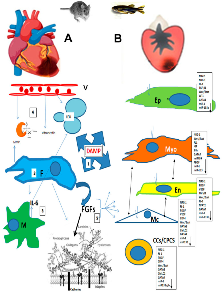
The adult human heart has a limited ability to regenerate after injury, leading to the formation of fibrotic scars and a subsequent loss of function. In fish, mice, and humans, cardiac remodeling after myocardial injury involves the activation of epicardial and endocardial cells, pericytes, stem cells, and fibroblasts. The heart's extracellular matrix (ECM) plays a significant role in the regeneration and recovery process. The epicardium, endocardium, and pericytes reactivate the embryonic program in response to ECM stimulation, which leads to epithelial-mesenchymal transition, cell migration, and differentiation. This review analyzes the role of ECM in guiding the differentiation or dedifferentiation and proliferation of heart components by comparing significant findings in a zebrafish model with those of mammals.
Keywords: extracellular matrix; heart; zebrafish.
Conflict of interest statement
The author declares no conflict of interest.
Figures
Similar articles
-
Lab-grown, 3D extracellular matrix particles improve cardiac function and morphology in myocardial ischemia.Am J Physiol Heart Circ Physiol. 2025 Feb 1;328(2):H221-H234. doi: 10.1152/ajpheart.00581.2024. Epub 2024 Dec 20. Am J Physiol Heart Circ Physiol. 2025. PMID: 39705507 Free PMC article.
-
Cardiac regeneration: Unraveling the complex network of intercellular crosstalk.Semin Cell Dev Biol. 2025 Jul;171:103619. doi: 10.1016/j.semcdb.2025.103619. Epub 2025 May 13. Semin Cell Dev Biol. 2025. PMID: 40367899 Review.
-
Cardiomyocyte maturation and its reversal during cardiac regeneration.Dev Dyn. 2024 Jan;253(1):8-27. doi: 10.1002/dvdy.557. Epub 2022 Dec 21. Dev Dyn. 2024. PMID: 36502296 Review.
-
Glucocorticoid Receptor Antagonism and Cardiomyocyte Regeneration Following Myocardial Infarction: A Systematic Review.Curr Probl Cardiol. 2023 Dec;48(12):101986. doi: 10.1016/j.cpcardiol.2023.101986. Epub 2023 Jul 20. Curr Probl Cardiol. 2023. PMID: 37481215
-
Activated cardiac fibroblasts are a primary source of high-molecular-weight hyaluronan production.Am J Physiol Cell Physiol. 2025 Mar 1;328(3):C939-C953. doi: 10.1152/ajpcell.00786.2024. Epub 2025 Jan 27. Am J Physiol Cell Physiol. 2025. PMID: 39871135 Free PMC article.
References
-
- Belviso I., Angelini F., Di Meglio F., Picchio V., Sacco A.M., Nocella C., Romano V., Nurzynska D., Frati G., Maiello C., et al. The Microenvironment of Decellularized Extracellular Matrix from Heart Failure Myocardium Alters the Balance between Angiogenic and Fibrotic Signals from Stromal Primitive Cells. Int. J. Mol. Sci. 2020;21:7903. doi: 10.3390/ijms21217903. - DOI - PMC - PubMed
Publication types
MeSH terms
Grants and funding
LinkOut - more resources
Full Text Sources
