. 2025 Jun 11;14(12):880.
doi: 10.3390/cells14120880.

Selective Blockade of Two Aquaporin Channels, AQP3 and AQP9, Impairs Human Leukocyte Migration

Affiliations

Selective Blockade of Two Aquaporin Channels, AQP3 and AQP9, Impairs Human Leukocyte Migration

Sabino Garra et al. Cells. .

Abstract

Peripheral blood leukocytes are able to migrate to the inflamed tissue, and to engulf and kill invading microbes. This requires rapid modifications of cell morphology and volume through fast movements of osmotic water into or out of the cell. In this process, membrane water channels, aquaporins (AQPs), are critical for cell shape changes as AQP-mediated water movement indirectly affects the cell cytoskeleton and, thereby, the signaling cascades. Recent studies have shown that the deletion or gating of two immune cell AQPs, AQP3 and AQP9, impairs inflammation and improves survival in microbial sepsis. Here, we assessed the expression and distribution of AQP3 and AQP9 in human leukocytes and investigated their involvement in the phagocytosis and killing of the Gram-negative pathogenic bacterium Klebsiella pneumoniae, and their role in lipopolysaccharide (LPS)-induced cell migration. By RT-qPCR, AQP3 mRNA was found in peripheral blood mononuclear cells (PBMCs) but it was undetectable in polymorphonuclear white blood cells (PMNs). AQP9 was found both in PBMCs and PMNs, particularly in neutrophil granulocytes. Immunofluorescence confirmed the AQP3 expression in monocytes and, to a lesser degree, in lymphocytes. AQP9 was expressed both in PBMCs and neutrophils. Specific inhibitors of AQP3 (DFP00173) and AQP9 (HTS13286 and RG100204) were used for bacterial phagocytosis and killing studies. No apparent involvement of individually blocked AQP3 or AQP9 was observed in the phagocytosis of K. pneumoniae by neutrophils or monocytes after 10, 30, or 60 min of bacterial infection. A significant impairment in the phagocytic capacity of monocytes but not neutrophils was observed only when both AQPs were inhibited simultaneously and when the infection lasted for 60 min. No impairment in bacterial clearance was found when AQP3 and AQP9 were individually or simultaneously blocked. PBMC migration was significantly impaired after exposure to the AQP9 blocker RG100204 in the presence or absence of LPS. The AQP3 inhibitor DFP00173 reduced PBMC migration only under LPS exposure. Neutrophil migration was considerably reduced in the presence of RG100204 regardless of whether there was an LPS challenge or not. Taken together, these results indicate critical but distinct involvements for AQP3 and AQP9 in leukocyte motility, while no roles are played in bacterial killing. Further studies are needed in order to understand the precise ways in which these two AQPs intervene during bacterial infections.

Keywords: LPS; aquaporin inhibitors; cell motility; host–bacteria relationship; inflammation; innate immunity; phagocytosis; white blood cells.


Conflict of interest statement

Author Michael Rützler was employed by the company Apoglyx AB. The remaining authors declare that the research was conducted in the absence of any commercial or financial relationships that could be construed as a potential conflict of interest.

Figures

Figure 1
Figure 1
Real-time quantitative PCR analysis of AQP1, 3, 5, and 9 mRNA expression in human white blood cells. PMNs, neutrophil granulocytes alone, and PBMCs were isolated from the peripheral blood of healthy donors (n = 5). The mRNA levels of the four AQPs were normalized against those of the housekeeper gene β-Actin. High levels of AQP9 mRNA are detected in polymorphonuclear leukocytes (A), especially in neutrophil granulocytes (B). The level of AQP9 transcript is considerably less abundant in PBMCs (C). AQP3 is expressed in PBMCs (C), while, in neutrophils, it is very weak or absent (A,B). AQP1 and AQP5 are barely appreciable or absent in all three WBC fractions (AC). Data are shown as mean ± SEM (n = 5). **, p < 0.01; ***, p < 0.001.
Figure 2
Figure 2
Immunofluorescence analysis of AQP3 and AQP9 in isolated human neutrophils. Neutrophils were isolated by immune-magnetic negative selection from the peripheral blood of healthy donors and analyzed using immunofluorescence with anti-AQP3 or anti-AQP9 polyclonal antibodies ((B) and (E), respectively). Cell nuclei were counterstained with DAPI ((A,D); blue fluorescence). (C) and (F) represent the merging of (A,B) and (D,E), respectively. Strong AQP9 immunofluorescence is seen in neutrophils, where it appears to be granular (arrows), and over the plasma membrane (E); green fluorescence). By contrast, neutrophils do not show any AQP3 immunoreactivity (B,C). Scale bars, 10 µm.
Figure 3
Figure 3
Immunofluorescence analysis of AQP3 and AQP9 in isolated human PBMCs. Isolated PBMCs were subjected to immunofluorescence (green) with anti-AQP3 (B,E) or anti-AQP9 (H) polyclonal antibodies. Cell nuclei were stained with DAPI ((A,D,G); blue fluorescence). Cells in (C), (F), and (I) are the merging of (A,B), (D,E), and (G,H), respectively. AQP3 immunoreactivity is high in monocytes (B,C), while it is barely appreciable in lymphocytes (E,F). Strong AQP9 immunofluorescence is instead seen in monocytes and lymphocytes both at the intracellular compartment (solid arrows) and over the plasma membrane (linear arrows) (H,I). Mc, monocyte; Lc, lymphocyte. Scale bars, 10 µm.
Figure 4
Figure 4
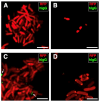
Human serum specimens are without antibodies to K. pneumoniae HA391. K. pneumoniae HA391 DsRed was incubated with the blood sera of three of the five subjects used for the study. An FITC-conjugated antibody against the human immunoglobulins G (hIgG; green fluorescence) was used to detect eventual anti-K. pneumoniae antibodies. (A) Negative control (absence of serum). (BD) K. pneumoniae HA391 DsRed incubated with each one of the three sera, separately. Arrows indicate unspecific spots located outside of bacterial bodies (C). Scale bars, 2 µm.
Figure 5
Figure 5
Phagocytosis of K. pneumoniae HA391 DsRed by blood neutrophils. Black and white micrograph of neutrophils in whole blood, stained with methylene blue. (A) Neutrophil from a blood sample incubated for 60 min with BHI medium. (B) Phagocytic-positive neutrophil from a blood sample infected for 60 min with K. pneumoniae HA391 DsRed in BHI medium. Arrows indicate the ingested bacteria. A distinct change in cell size and morphology is noted compared to the uninfected neutrophil shown in (A). n, nucleus. Scale bars, 2 µm.
Figure 6
Figure 6
Flow cytometry analysis of neutrophil phagocytosis of K. pneumoniae after AQP3 and/or AQP9 inhibition. Whole blood was exposed to K. pneumoniae HA391 DsRed, or BHI control medium (with or without vehicle) for 10, 30, or 60 min. After red blood cells lysis and washout, flow cytometry analysis of neutrophils was conducted by selection through the CD66b marker. Percentages of RFP-positive neutrophils were calculated. (A) Basal condition. Blood sample exposed to the medium alone for 60 min (no infection). (BD) Blood samples infected with K. pneumoniae for 10, 30, or 60 min, respectively. About one third of neutrophils are infected after 60 min of exposure to K. pneumoniae HA391 DsRed (D). (EG) Percentage of RFP-positive in the different control and experimental conditions (inset), following 10, 30, or 60 min of infection, respectively. There are no statistically significant differences among the different conditions. Vehicle, 1% DMSO. Percentages are expressed as mean ± SEM (n = 5). a.u., arbitrary units.
Figure 7
Figure 7
Flow cytometry analysis of monocyte phagocytosis of K. pneumoniae after AQP3 and/or AQP9 inhibition. Whole blood was infected (or not) with BHI medium containing K. pneumoniae. After washout and red blood cell lysis, flow cytometry analysis of monocytes was conducted by selection through the CD14 marker. Percentages of phagocytic-positive monocytes were calculated through the signal of the internalized red fluorescent protein (RFP). (A) Basal condition. Blood sample exposed to the medium alone for 60 min (no infection). (BD) Blood samples infected with K. pneumoniae for 10, 30, or 60 min, respectively. More than 40% of monocytes are infected after 60 min exposure to the bacterial strain (D). (EG) Percentage of phagocytic-positive monocytes (RFP+) in the different control and experimental conditions (see inset) following 10, 30, or 60 min of infection, respectively. The only statistically significant difference is seen between the control with no vehicle and the experimental condition where the two inhibitors, DFP00173 and HTS13286, were both added. Vehicle, 1% DMSO. Percentages are expressed as mean ± SEM (n = 5). *, p < 0.05.
Figure 8
Figure 8
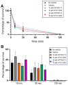
Inhibition of AQP3 and AQP9 does not impair killing of K. pneumoniae by WBCs. Whole blood samples were exposed to the BHI medium containing K. pneumoniae for up to 120 min and intracellular bacteria were quantified by lysis, serial dilution, and viable counting on LB agar plates (see Materials and Methods for details). (A) Survival curves of K. pneumoniae inoculated in whole blood samples and evaluated after 10, 30, and 120 min of incubation in absence or presence of vehicle (1% DMSO) and AQP3/AQP9 selective inhibitor. In all conditions, all inoculated bacteria are almost totally eliminated within 120 min. (B) Percentages of bacterial survival in presence or absence of the AQP3 or AQP9 inhibitor after 10, 30, or 120 min of exposure to whole blood. Both DFP00173 and HTS13286 do not impair the clearance efficacy of leukocytes. Percentages are expressed as mean ± SEM (n = 5).
Figure 9
Figure 9
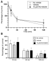
Killing of K. pneumoniae by PMNs is not impaired by the AQP9 inhibitor RG100204. Isolated PMNs were infected with K. pneumoniae for up to 120 min and surviving intracellular bacteria were quantified by lysis, serial dilution, and viable counting on LB agar plates (see Materials and Methods for details). (A) Survival curve of K. pneumoniae inoculated in PMN samples and analyzed after 10, 30, and 120 min of incubation in absence or presence of vehicle or 10 µM RG100204. In all three conditions, after 120 min of incubation, nearly 90% of bacteria are resolved by PMNs. (B) Percentages of bacterial survival in presence or absence (see inset for conditions) of the AQP9-selective inhibitor RG100204 after 10, 30, or 120 min of killing by PMNs. RG100204 does not seem to affect the killing capacity of PMNs. Percentages are expressed as mean ± SEM (n = 5).
Figure 10
Figure 10
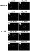
Effect of RG100204 on LPS-induced cell migration of human PMNs. Representative micrographs showing the PMNs that had migrated (white spots) across the transwell filter after 16 h with (panels (CJ)) or without (panels (A,B)) LPS challenging in absence (vehicle, 0.5% DMSO; panel (C)) or presence of a series of doses of the AQP9 inhibitor RG100204 (RG; panels (DJ)). Scale bars, 150 µm.
Figure 11
Figure 11
The AQP9-selective inhibitor RG100204 reduces neutrophil migration. Migration analysis of isolated whole PMNs after 16 h with or without LPS challenge, and in presence or absence (vehicle alone, 0.5% DMSO; V) of serial concentrations of RG100204 (RG). Transmigrated PMNs are expressed as percentage, compared to the number of PMNs migrating without LPS stimulation and in the presence of the vehicle alone. Percentages are expressed as mean ± SEM (n = 5; 6 fields/condition). ** p < 0.01; *** p < 0.001.
Figure 12
Figure 12
Selective inhibition of both AQP3 and AQP9 reduces PBMC migration. Migration analysis of isolated PBMCs after 16 h with or without LPS challenge and in absence (vehicle alone, 0.5% DMSO; V) or presence of 10 µM DFP00173 (DFP) or 10 µM RG100204 (RG). Migrated PBMCs are expressed as percentage of PBMCs migrating without LPS stimulation and in presence of vehicle alone. Percentages are expressed as mean ± SEM (n = 5; 10 fields/condition). ** p < 0.01; *** p < 0.001.

Similar articles

Cited by

References

    1. Fenteany G., Glogauer M. Cytoskeletal remodeling in leukocyte function. Curr. Opin. Hematol. 2004;11:15–24. doi: 10.1097/00062752-200401000-00004. - DOI - PubMed
    1. Papadopoulos M.C., Saadoun S., Verkman A.S. Aquaporins and cell migration. Pflug. Arch. 2008;456:693–700. doi: 10.1007/s00424-007-0357-5. - DOI - PMC - PubMed
    1. Loitto V.M., Forslund T., Sundqvist T., Magnusson K.E., Gustafsson M. Neutrophil leukocyte motility requires directed water influx. J. Leukoc. Biol. 2002;71:212–222. doi: 10.1189/jlb.71.2.212. - DOI - PubMed
    1. Zhu N., Feng X., He C., Gao H., Yang L., Ma Q., Guo L., Qiao Y., Yang H., Ma T. Defective macrophage function in aquaporin-3 deficiency. FASEB J. 2011;25:4233–4239. doi: 10.1096/fj.11-182808. - DOI - PubMed
    1. Holm A., Karlsson T., Vikstrom E. Pseudomonas aeruginosa lasI/rhlI quorum sensing genes promote phagocytosis and aquaporin 9 redistribution to the leading and trailing regions in macrophages. Front. Microbiol. 2015;6:915. doi: 10.3389/fmicb.2015.00915. - DOI - PMC - PubMed

MeSH terms

LinkOut - more resources