. 2025 Jun 12;14(12):888.
doi: 10.3390/cells14120888.

Wnt/PKC Signaling Inhibits Sensory Hair Cell Formation in the Developing Mammalian Cochlea

Affiliations

Wnt/PKC Signaling Inhibits Sensory Hair Cell Formation in the Developing Mammalian Cochlea

Joanna F Mulvaney et al. Cells. .

Abstract

The establishment of cell fate and boundaries between cell types is an essential step in development and organogenesis. In the mammalian cochlea, a distinct boundary exists between a medial region of non-sensory cells and a lateral region of sensory cells. We report that Wnt4 and sFRP2 act in combination to modulate the sensory cell differentiation of the organ of Corti. The hair cell inhibitory effects of Wnt4 in the inner ear are mediated through the activation of the non-canonical Wnt/Calcium/PKC pathway. We show that Wnt4 stimulates the activation of PKC in the cochlea, and that the inhibition of PKC rescues the ectopic Wnt4 activity phenotype. Finally, we demonstrate that modification at a PKC target site on Atoh1 diminishes its ability to induce hair cell formation. Ultimately, we identify a new Wnt/Calcium/PKC non-canonical signaling pathway that is involved in proper hair cell and organ of Corti formation in the developing mammalian cochlea.

Keywords: cochlea; genes; hair cell; hearing; mouse.


Conflict of interest statement

The authors declare no conflicts of interest. The funders had no role in the design of this study; in the collection, analyses, or interpretation of data; in the writing of the manuscript; or in the decision to publish the results.

Figures

Figure 1
Figure 1
Wnt4 is expressed in the developing cochlea. (A) Semi-thin cross-section through the basal turn of an E17 cochlea stained with hematoxylin and eosin (H&E) to illustrate the composition of a wild-type developing cochlear duct. A single IHC (arrowhead) and three OHCs (three arrows) can be identified in the organ of Corti (OC). In all cross-sections, medial (modiolar, Med) is toward the left, and lateral (strial, Lat) is toward the right. RM: Reissner’s membrane. (B) In situ hybridization for Wnt4 in a cross-section through the basal turn of an E13 cochlea. Wnt4 is expressed in the medial half of the roof of the cochlear duct. The expression of Wnt4 is less than 50 µm from the prospective OC (indicated by an asterisk). (C) Cross-section through the basal turn of an E14.5 cochlea. Wnt4 expression persists in the medial region of the roof of the cochlear duct. (D) Cross-section through the basal turn of an E16.5 cochlea. Wnt4 expression is restricted to cells at the medial edge of the developing Reissner’s membrane. The scale bar represents 50 µm.
Figure 2
Figure 2
Wnt4 is necessary for limiting the number of inner hair cells in vivo. (A) Whole mount surface view of a P0 cochlea (mid-base) from a wild-type mouse with the normal pattern of one row of IHCs and three rows of OHCs labeled with anti-Myosin6. (B) Surface view of the mid-base of a P0 Wnt4−/− cochlea. In addition to the one row of IHCs and three rows of OHCs, extra IHCs (arrows) are present along the length of the OC. (A’) and (B’) Zoomed in confocal images of (A) and (B), respectively. (C) Cross-section of the OC from a P0 wild-type mouse. The normal pattern of one IHC and three OHCs (1, 2, 3) is present. (D) Cross-section of the OC from a P0 Wnt4−/− cochlea. Two IHCs (asterisks) and three OHCs (1, 2, 3) are present. (E) Average number of ectopic IHCs in P0 cochleae from WT, Wnt4+/−, and Wnt4−/− mice. A significant increase in the number of IHCs was present in Wnt4−/− cochlea as compared to wild-type littermates (Welch’s two-tailed T-test, n = 9 for each genotype; p = 0.0003). Asterisks indicate statistical significance (*). (F) Average number of OHCs in cochleae from P0 WT, Wnt4+/−, and Wnt4−/− mice. There was no significant (NS) change in the number of OHCs in cochleae from any of the three genotypes in a 100 µm region along the mid-base of the cochleae (ANOVA; p = 0.4, F = 0.87). (G) There was no significant difference in the lengths of the wild type and Wnt4−/− cochleae, indicating that there is no convergence and extension phenotype (ANOVA, p = 0.18, F = 2.3). Error bars indicate SEM. Scale bar in (A) and (A’) represents 20 µm, scale bar in (C) represents 10 µm.
Figure 3
Figure 3
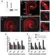
Wnt4 inhibits hair cell formation in vitro. (A) Western blot showing that the Wnt4-conditioned media (Wnt4-CM) contained Wnt4 protein. (B) E13 cochlear explant cultured for 6 days in vitro in control-conditioned media (Control-CM). A single row of IHCs and three rows of OHCs (labeled with anti-Myosin6) are present. Percentages indicate the percentage distance from the base of the OC. (C) Explant treated with Wnt4-CM beginning at E13. The number of HCs is clearly reduced, especially in the apical region. (D) Treatment with Wnt4-CM beginning at E15. (E) Wnt4 inhibits IHC formation. (B’), (C’), and (D’) are zoomed-in confocal images of (B), (C), and (D), respectively, from the apical (approximately 75%) region of the explant. The number of IHCs was determined for a 100 µm length of the OC at the positions indicated. A significant reduction in the number of cells that developed as IHCs was observed in cochleae treated with 1× or 10× Wnt4-CM at each position (Student’s test, two-tailed, n = 6 for each treatment condition. * p < 0.02, ** p < 0.002). (F) Wnt4 inhibits OHC formation. Wnt4-CM resulted in a significant reduction in the number of cells that developed as OHCs along the length of the cochlea (n = 6; * p < 0.02, ** p < 0.0007). Error bars indicate SEM. Scale bars represent 50 µm.
Figure 4
Figure 4
Wnt antagonist sFRP2 is expressed contemporaneously with Wnt4 and has the reverse effect of Wnt4. (A) Cross-section through the cochlear duct at E13 illustrating the expression of Wnt4 (red, arrow) protein by immunohistochemistry. The asterisk indicates the prospective organ of Corti. (B) Similar cross-section as in A, showing sFRP2 expression by immunohistochemistry. sFRP2 (red, arrow) is expressed on the lateral edge of the cochlear duct—lateral to the area where the OC (*) will develop. (C) Surface view of the mid-base of a control cochlear explant culture established on E13 and maintained for 6 days in vitro. A single row of IHCs and three to four rows of OHCs are labeled with anti-Myosin6 (red). (D) Similar view as in C from an explant established on E13 and treated with 50 µg/mL sFRP2. An almost continuous second row of IHCs is present. Scale bars represent 50 μm.
Figure 5
Figure 5
Calcium chelation induces ectopic IHCs in the developing organ of Corti. (A) Surface view of the mid-base of an explant culture established at E13 and maintained in control DMSO media for 6 days, the typical three rows of OHCs and one row of IHCs are present. (B) Surface view of the mid base of an explant culture established on day E13 and maintained in media containing 20 µM of the calcium chelator BAPTA-AM. There is an increase in the number of cells that develop into IHCs. (A) and (B) show the mid-base of the cochlea; hair cells are labeled with anti-Myosin6. (C) Quantification of the increase in IHC and OHC number in cultures treated with BAPTA-AM. Cell counts were taken at 25%, 50%, and 75% of the length of the explant. (*) p < 0.05; (**) p < 0.001; 3 independent experiments for each treatment condition. Bars indicate SEM. The scale bar represents 50 μm.
Figure 6
Figure 6
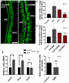
Inhibition of PKC induces the formation of ectopic IHCs in the developing organ of Corti. (A) An explant organ culture established at E13 and maintained in control media for 6 days. The typical three rows of OHCs and one row of IHCs are present. (B) An explant culture established at E13 treated with the PKC inhibitor BIM I (2 µM). Supernumerary IHCs have differentiated. Images (A) and (B) show the mid-base of the cochlear cultures, HCs labeled with anti-Myosin7a. (C) Quantification of the increase in IHCs in response to the activation of PKC at different stages of development. BIM I was added to explant cultures at E13, E14, and E15. Relative to control E13 explants cultured in DMSO (n = 3), there was a significant increase in the number of IHCs when cultures were exposed to BIM I beginning on E13 (n = 3) and 14 (n = 3); this effect was abolished by E15 (n = 3). Error bars represent SEM, * p < 0.05. (D) Quantification of the increase in IHCs in response to the inhibition of PKC. Two highly selective PKC inhibitors, BIM I (n = 3) or Gö6983 (n = 3), significantly increased the number of IHCs compared to explants cultured in DMSO (n = 3). BIM V, used as a negative control, had no significant effect. * p < 0.01. (E) Quantification of Atoh1, Hes1, and Hes5 expression levels in control and BIM I treated cochlear explant cultures by real-time quantitative PCR. In BIM I treated cultures, Atoh1 was expressed 2.02-fold more than DMSO treated control cultures after 48 h of treatment, p = 0.007. Hes1 was elevated 1.97-fold, p = 0.005. Hes5 expression levels were unchanged (fold change was 0.97). Control n = 3. BIM I treated n = 3. Asterisks (*) denote statistical significance. The scale bar represents 50 µm. (F) Quantification of BrdU-positive hair cells (p = 0.5) in explant cultures treated with BIM I, concurrent with the expansion in the number of inner hair cells. n = 3, error bars indicate SEM, NS = no significance.
Figure 7
Figure 7
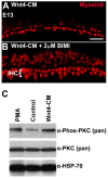
PKC, activated by Wnt4, modulates cell fate. (A) An image of a region in the mid-base of an explant established and treated with Wnt4-CM beginning at E13. As illustrated in Figure 2, the number of HCs is reduced in the presence of Wnt4. (B) Explant from mid-base treated with Wnt4-CM and the PKC inhibitor BIM I (2 µM). Inhibition of PKC not only blocks the inhibitory effect of Wnt4 but results in the formation of extra IHCs, as was observed in the presence of BIM I alone. (C) Wnt4 activates PKC in cochlear tissue. Phosphorylation of PKC was determined in E14 cochleae, untreated, treated with the PKC activator PMA, or treated with Wnt4-CM. The total level of PKC, as assayed using the pan-PKC antibody, was unchanged between the three treatments; however, the levels of phospho-PKC were increased in cochleae treated with PMA or Wnt4-CM. HSP-70 levels were assayed as a loading control. The scale bar represents 50 µm.
Figure 8
Figure 8
Atoh1 function is ablated by phosphorylation of a PKC target site. (A) Cochlear explant culture established on E13 after 6 days in vitro. Hair cells are labeled with anti-Myosin6 (red). Non-sensory epithelial cells located medial to the sensory epithelium (SE) have been transfected with the Atoh1(S146D).EGFP mutant. Expression of Atoh1(S146D) induces the formation of ectopic hair cells (yellow cells). (B) Cochlear explant transfected with the Atoh1(T197D).EGFP mutant showed no induced hair cells. Note that none of the green cells are positive for expression of Myosin6 (red). (CF) Overexpression of Atoh1(T197D).EGFP results in Atoh1 protein formation in HEK293 cells. (C) Two HEK293 cells marked by DAPI (blue), one of which has been transfected with Atoh1(T197D).EGFP (green). The transfected cell is positive for Atoh1 protein (red). (DF) show individual channels. Scale bars represent 10 µm.

References

    1. Driver E.C., Kelley M.W. Development of the cochlea. Development. 2020;147:dev162263. doi: 10.1242/dev.162263. - DOI - PMC - PubMed
    1. Kelley M.W. Regulation of cell fate in the sensory epithelia of the inner ear. Nat. Rev. Neurosci. 2006;7:837–849. doi: 10.1038/nrn1987. - DOI - PubMed
    1. Kelley M.W., Driver E.C., Puligilla C. Regulation of cell fate and patterning in the developing mammalian cochlea. Curr. Opin. Otolaryngol. Head Neck Surg. 2009;17:381–387. doi: 10.1097/MOO.0b013e3283303347. - DOI - PMC - PubMed
    1. Groves A.K., Fekete D.M. Shaping sound in space: The regulation of inner ear patterning. Development. 2012;139:245–257. doi: 10.1242/dev.067074. - DOI - PMC - PubMed
    1. Webber J.L., Garcia-Anoveros J. Precision patterning: How inner hair cells “hop” to it. Sci. Adv. 2023;9:eadg8662. doi: 10.1126/sciadv.adg8662. - DOI - PMC - PubMed

LinkOut - more resources