Review
. 2025 Jun 18;14(12):918.
doi: 10.3390/cells14120918.

Cellular and Molecular Interactions in CNS Injury: The Role of Immune Cells and Inflammatory Responses in Damage and Repair

Affiliations
Review

Cellular and Molecular Interactions in CNS Injury: The Role of Immune Cells and Inflammatory Responses in Damage and Repair

Jai Chand Patel et al. Cells. .

Abstract

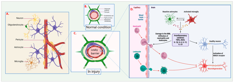
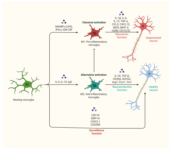
The central nervous system (CNS) is highly susceptible to damage due to its limited ability to regenerate. Injuries to the CNS, whether from trauma, ischemia, or neurodegenerative diseases, disrupt both cellular and vascular structures, leading to immediate (primary) and subsequent (secondary) damage. Primary damage involves the physical disruption of cells and blood vessels, weakening the blood-brain barrier (BBB) and triggering excitotoxicity and calcium overload. Secondary damage develops over hours to days and is marked by ionic imbalance, mitochondrial dysfunction, oxidative stress, and chronic inflammation, which further aggravates tissue damage. Inflammation plays a dual role: acute inflammation helps in repair, while chronic inflammation accelerates neurodegeneration. Microglia and astrocytes play key roles in this inflammatory response, with M1-like microglia promoting pro-inflammatory responses and M2-like microglia supporting anti-inflammatory and repair processes. Neurodegenerative diseases are characterized by the accumulation of misfolded proteins such as Tau, amyloid-beta, TDP-43, and α-synuclein, which impair cellular function and lead to neuronal loss. Neurodegenerative diseases are characterized by the accumulation of misfolded proteins and influenced by genetic risk factors (e.g., APOE4, TARDBP). Despite the CNS's limited regenerative abilities, processes like synaptogenesis, neurogenesis, axonal regeneration, and remyelination offer potential for recovery. Therapeutic approaches aim to target inflammatory pathways, enhance repair mechanisms, and develop neuroprotective treatments to counter excitotoxicity, oxidative stress, and apoptosis. Advances in stem cell therapy, gene therapy, and personalized medicine hold promise for improving outcomes. Future research should focus on combining strategies, utilizing advanced technologies, and conducting translational studies to bridge the gap between preclinical research and clinical application. By better understanding and leveraging the complex processes of CNS injury and repair, researchers hope to develop effective therapies to restore function and enhance the quality of life for individuals with CNS disorders.

Keywords: CNS injury; excitotoxicity; inflammation; microglia; neurodegenerative diseases; neurogenesis.


Conflict of interest statement

The authors declare no conflicts of interest.

Figures

Figure 1
Figure 1
Cellular and molecular responses in the central nervous system (CNS) under normal and injured conditions: (A) Overview of major CNS cell types: Neurons, oligodendrocytes, pericytes, astrocytes, and microglia. (B) Normal conditions: Under typical physiological circumstances, astrocytes and pericytes help maintain the integrity of the blood–brain barrier (BBB), keeping its membrane structure intact. (C) Disrupted barrier in injury: Injury to the BBB results in its compromise, causing the barrier to become leaky and permitting harmful substances and cells to infiltrate the CNS. (D) Capillary and BBB components: Depicts the structure of capillaries, the blood–brain barrier, neuroglial cells, and leukocytes. In response to injury, astrocytes become reactive, and microglia are activated. This activation contributes to neural tissue damage, the infiltration of neutrophils and other leukocytes, and the release of pro-inflammatory cytokines such as TNF-α, IL-1β, IL-6, IL-12, and IL-23. The illustration contrasts a healthy neuron with intact NMDA receptors against the neurodegeneration resulting from inflammatory and cytotoxic processes following injury.
Figure 2
Figure 2
Activation states of microglia and their functional outcomes: Classical activation (M1 phenotype): Induced by MAMPs (e.g., LPS), IFN-γ, and GM-CSF. This state is characterized by the release of pro-inflammatory cytokines and mediators, including IL-1β, IL-6, IL-12, TNF-α, CCL2, CXCL10, iNOS, MHC II, CD86, and CD16/32. M1 activation is associated with neurotoxic effects, contributing to neuronal damage and degeneration. Alternative activation (M2 phenotype): This represents an anti-inflammatory response by microglia. In this state, microglia secrete anti-inflammatory cytokines such as IL-10 and TGF-β and express markers including Arg1 and Ym1, etc. M2 activation supports neuroprotective functions, helping to maintain healthy neurons. Additionally, it plays a role in surveillance activities mediated by CSF1R, SIRP1A, CX3CL1, and CD200R.
Figure 3
Figure 3
Microglial and astrocytic responses in normal and pathological states. Normal state: In the healthy CNS, microglia and astrocytes work together to maintain homeostasis. Neurons remain functional and undamaged. In response to inflammatory signals, microglia become activated, and astrocytes become reactive. This activation leads to neuronal degeneration and disrupts normal neural function. Microglia exhibit enhanced actin cytoskeleton recognition, promoting cell survival, increased phagocytosis, and improved clearance of amyloid-beta (Aβ). This results in a reduction in Aβ accumulation. However, pathological features such as tau protein tangles, disrupted microtubules, amyloid-beta plaques, neurofibrillary tangles, and necrotic tangles emerge, all of which contribute to the progression of neurodegenerative diseases.

Similar articles

Cited by

References

    1. Tam R.Y., Fuehrmann T., Mitrousis N., Shoichet M.S. Regenerative Therapies for Central Nervous System Diseases: A Biomaterials Approach. Neuropsychopharmacology. 2014;39:169–188. doi: 10.1038/npp.2013.237. - DOI - PMC - PubMed
    1. Varadarajan S.G., Hunyara J.L., Hamilton N.R., Kolodkin A.L., Huberman A.D. Central nervous system regeneration. Cell. 2022;185:77–94. doi: 10.1016/j.cell.2021.10.029. - DOI - PMC - PubMed
    1. Brett B.L., Gardner R.C., Godbout J., Dams-O’Connor K., Keene C.D. Traumatic Brain Injury and Risk of Neurodegenerative Disorder. Biol. Psychiatry. 2022;91:498–507. doi: 10.1016/j.biopsych.2021.05.025. - DOI - PMC - PubMed
    1. Logsdon A.F., Lucke-Wold B.P., Turner R.C., Huber J.D., Rosen C.L., Simpkins J.W. Role of Microvascular Disruption in Brain Damage from Traumatic Brain Injury. Compr. Physiol. 2015;5:1147–1160. doi: 10.1002/j.2040-4603.2015.tb00640.x. - DOI - PMC - PubMed
    1. Price L., Wilson C., Grant G. Blood-Brain Barrier Pathophysiology Following Traumatic Brain Injury. CRC Press; Boca Raton, FL, USA: 2016. - PubMed

MeSH terms

LinkOut - more resources