Case Reports
. 2025 May 27;13(6):170.
doi: 10.3390/diseases13060170.

Intraosseous Pneumatocysts of the Scapula Mimicking Bone Tumors: A Report of Two Rare Cases Along with Elucidation of Their Etiology

Affiliations
Case Reports

Intraosseous Pneumatocysts of the Scapula Mimicking Bone Tumors: A Report of Two Rare Cases Along with Elucidation of Their Etiology

Jiro Ichikawa et al. Diseases. .

Abstract

Background/objectives: Pneumatocysts, characterized by gas-filled cavities, are commonly found in the spine and pelvis but are rarely observed in the scapula. In this report, we describe two rare cases of scapular pneumatocysts mimicking bone tumors and exhibiting different image findings.

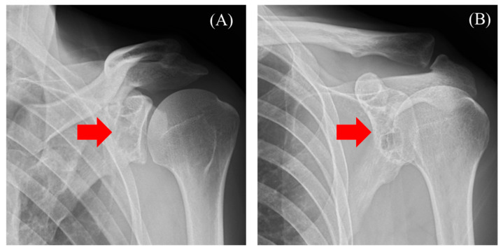
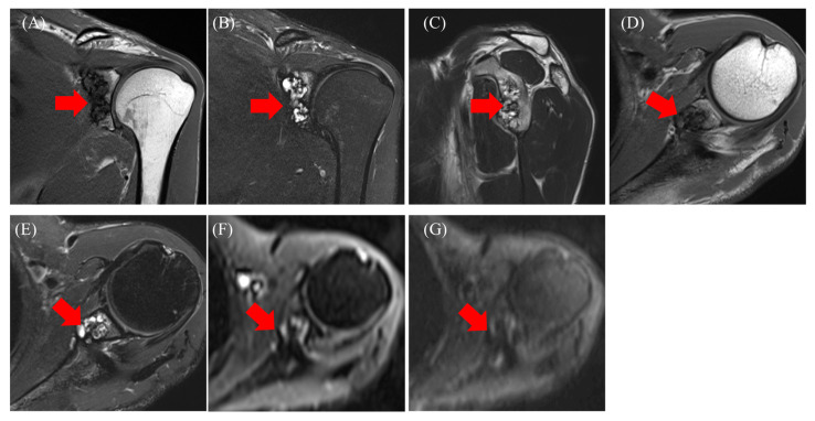
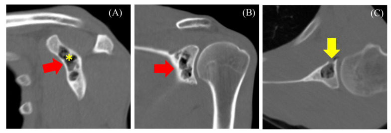
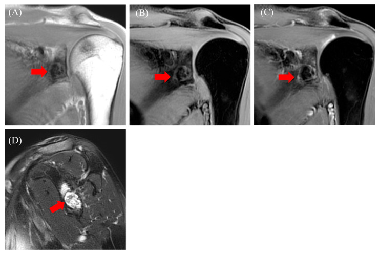
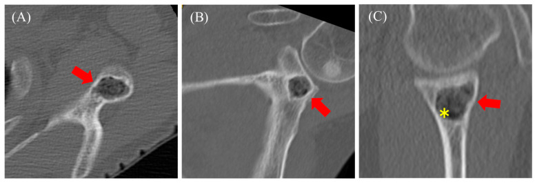
Case report: Case 1. A 47-year-old man who presented with neck pain underwent radiography, followed by magnetic resonance imaging (MRI). MRI showed heterogeneity with low and high signals on fat-suppressed T2-weighted images, suggestive of enchondroma or fibrous dysplasia (FD). However, preoperative computed tomography (CT) revealed gas-filled cavities within the tumor, in continuity with the shoulder joint, confirming the diagnosis of a pneumatocyst.

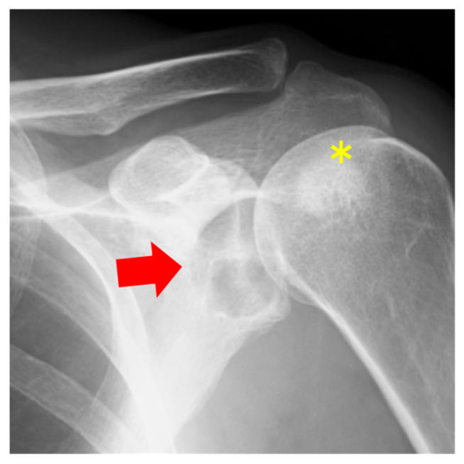
Case 2: A 58-year-old woman who presented with neck pain underwent similar examinations to Case 1. MRI showed homogeneity with high signals on fat-suppressed T2-weighted images, leading to a suspicion of solitary bone cysts and FD. Preoperative CT revealed gas-filled cavities within the tumor, but no continuity with the joint, leading to the diagnosis of a pneumatocyst. While the exact etiology of pneumatocysts remains unclear, two potential causes are as follows: (i) gas migration from the joint to the bone, and (ii) gas replacement in cystic tumors. Thus, CT is particularly valuable in confirming the presence of gas-filled cavities and aiding in diagnosis.

Conclusions: This report highlights two extremely rare cases of scapular pneumatocysts, reflecting two potential etiologies. The utility of CT in the diagnosis of pneumatocyst has been clarified.

Keywords: computed tomography; differential diagnosis; magnetic resonance imaging; pneumatocyst.


Conflict of interest statement

The authors declare no conflicts of interest.

Figures

Figure 1
Figure 1
Radiography reveals a radiolucent area with a sclerotic margin ((A) red arrow) and ((B) red arrow).
Figure 2
Figure 2
Magnetic resonance imaging: T1-weighted images (A,D), T2-weighted (C), fat-suppressed T2-weighted images (B,E), diffusion-weighted images b = 0 (F), b = 800 (G). agnetic resonance imaging: T1-weighted images ((A,D) red arrow), T2-weighted ((C) red arrow), fat-suppressed T2-weighted images ((B,E) red arrow), diffusion-weighted images b = 0 ((F) red arrow), b = 800 ((G) red arrow).
Figure 3
Figure 3
Computed tomography reveals a lobulated translucent image with a sclerotic margin and scattered air density within the lesion ((AC) red arrow). The asterisk indicates the location where CT value was measured. Bone defects in certain areas suggest communication with the joint ((C), yellow arrow).
Figure 4
Figure 4
Radiography shows a radiolucent area with a sclerotic margin (red arrow) and sclerotic regions of the humeral head (yellow asterick).
Figure 5
Figure 5
Magnetic resonance imaging: T1-weighted images ((A), red arrow), fat-suppressed T1-weighted images ((B), red arrow), fat-suppressed T1-weighted images with contrast ((C), red arrow), and fat-suppressed T2-weighted images ((D), red arrow).
Figure 6
Figure 6
Computed tomography reveals a lobulated translucent image with a sclerotic margin and faint air density within the lesion ((AC) red arrow). The asterisk indicates the location where CT value was measured.

Similar articles

References

    1. Oehler E., Valour F., Pascart T. Intraosseous pneumatocysts: A case based review. Clin. Rheumatol. 2013;32:129–134. doi: 10.1007/s10067-012-2077-2. - DOI - PubMed
    1. Garg K., Dash A., Aggarwal A., Duetzmann S. Vertebral Pneumatocyst—A Systematic Review. World Neurosurg. 2021;151:77–86. doi: 10.1016/j.wneu.2021.04.092. - DOI - PubMed
    1. Southi J.T., Estephan T., Mitchelle A., Loneragan R. Intraosseous pneumatocyst of the scapula: A case report. Radiol. Case Rep. 2024;19:4599–4603. doi: 10.1016/j.radcr.2024.07.029. - DOI - PMC - PubMed
    1. Kamba M., Ohuchi Y., Ogawa T., Ohata R. Intraosseous pneumatocyst of the scapula. Br. J. Radiol. 2000;73:658–660. doi: 10.1259/bjr.73.870.10911791. - DOI - PubMed
    1. Ramirez H., Jr., Blatt E.S., Cable H.F., McComb B.L., Zornoza J., Hibri N.S. Intraosseous pneumatocysts of the ilium. Findings on radiographs and CT scans. Radiology. 1984;150:503–505. doi: 10.1148/radiology.150.2.6691108. - DOI - PubMed

Publication types

LinkOut - more resources