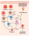
Erythrodermic Psoriasis in the Context of Emerging Triggers: Insights into Dupilumab-Associated and COVID-19-Induced Psoriatic Disease
- PMID: 40558675
- PMCID: PMC12192188
- DOI: 10.3390/dermatopathology12020017
Erythrodermic Psoriasis in the Context of Emerging Triggers: Insights into Dupilumab-Associated and COVID-19-Induced Psoriatic Disease
Abstract
Psoriasis is a chronic immune-mediated inflammatory skin disorder characterized by keratinocyte hyperproliferation, impaired epidermal barrier function, and immune dysregulation. The Th17/IL-23 axis plays a central role in its pathogenesis, promoting the production of key pro-inflammatory cytokines such as IL-17, IL-23, and TNF-α, which sustain chronic inflammation and epidermal remodeling. Emerging evidence suggests that SARS-CoV-2 may trigger new-onset or exacerbate existing psoriasis, likely through viral protein-induced activation of toll-like receptors (TLR2 and TLR4). This leads to NF-κB activation, cytokine release, and enhanced Th17 responses, disrupting immune homeostasis. Erythrodermic psoriasis (EP), a rare and severe variant, presents with generalized erythema and desquamation, often accompanied by systemic complications, including infection, electrolyte imbalance, and hemodynamic instability. In a murine model of SARS-CoV-2 infection, we found notable cutaneous changes: dermal collagen deposition, hair follicle destruction, and subcutaneous adipose loss. Parallel findings were seen in a rare clinical case (only the third reported case) of EP in a patient with refractory psoriasis, who developed erythroderma after off-label initiation of dupilumab therapy. The patient's histopathology closely mirrored the changes seen in the SARS-CoV-2 model. Histological evaluations also reveal similarities between psoriasis flare-ups following dupilumab treatment and cutaneous manifestations of COVID-19, suggesting a shared inflammatory pathway, potentially mediated by heightened type 1 and type 17 responses. This overlap raises the possibility of a latent connection between SARS-CoV-2 infection and increased psoriasis severity. Since the introduction of COVID-19 vaccines, sporadic cases of EP have been reported post-vaccination. Although rare, these events imply that vaccine-induced immune modulation may influence psoriasis activity. Our findings highlight a convergence of inflammatory mediators-including IL-1, IL-6, IL-17, TNF-α, TLRs, and NF-κB-across three triggers: SARS-CoV-2, vaccination, and dupilumab. Further mechanistic studies are essential to clarify these relationships and guide management in complex psoriasis cases.
Keywords: COVID-19; IL-4; SARS-CoV-2; T-cell; dupilumab; erythrodermic psoriasis; monoclonal antibodies; psoriasis.
Conflict of interest statement
The authors declare no conflicts of interest.
Figures


Similar articles
-
Systemic pharmacological treatments for chronic plaque psoriasis: a network meta-analysis.Cochrane Database Syst Rev. 2021 Apr 19;4(4):CD011535. doi: 10.1002/14651858.CD011535.pub4. Cochrane Database Syst Rev. 2021. Update in: Cochrane Database Syst Rev. 2022 May 23;5:CD011535. doi: 10.1002/14651858.CD011535.pub5. PMID: 33871055 Free PMC article. Updated.
-
Systemic pharmacological treatments for chronic plaque psoriasis: a network meta-analysis.Cochrane Database Syst Rev. 2017 Dec 22;12(12):CD011535. doi: 10.1002/14651858.CD011535.pub2. Cochrane Database Syst Rev. 2017. Update in: Cochrane Database Syst Rev. 2020 Jan 9;1:CD011535. doi: 10.1002/14651858.CD011535.pub3. PMID: 29271481 Free PMC article. Updated.
-
Antibody tests for identification of current and past infection with SARS-CoV-2.Cochrane Database Syst Rev. 2022 Nov 17;11(11):CD013652. doi: 10.1002/14651858.CD013652.pub2. Cochrane Database Syst Rev. 2022. PMID: 36394900 Free PMC article.
-
Systemic treatments for eczema: a network meta-analysis.Cochrane Database Syst Rev. 2020 Sep 14;9(9):CD013206. doi: 10.1002/14651858.CD013206.pub2. Cochrane Database Syst Rev. 2020. PMID: 32927498 Free PMC article.
-
Signs and symptoms to determine if a patient presenting in primary care or hospital outpatient settings has COVID-19.Cochrane Database Syst Rev. 2022 May 20;5(5):CD013665. doi: 10.1002/14651858.CD013665.pub3. Cochrane Database Syst Rev. 2022. PMID: 35593186 Free PMC article.
References
LinkOut - more resources
Full Text Sources
Miscellaneous

