. 2025 May 30;11(6):419.
doi: 10.3390/gels11060419.

Constructive Neuroengineering of Crossing Multi-Neurite Wiring Using Modifiable Agarose Gel Platforms

Affiliations

Constructive Neuroengineering of Crossing Multi-Neurite Wiring Using Modifiable Agarose Gel Platforms

Soya Hagiwara et al. Gels. .

Abstract

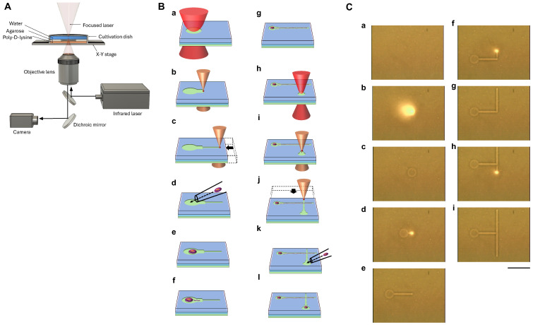
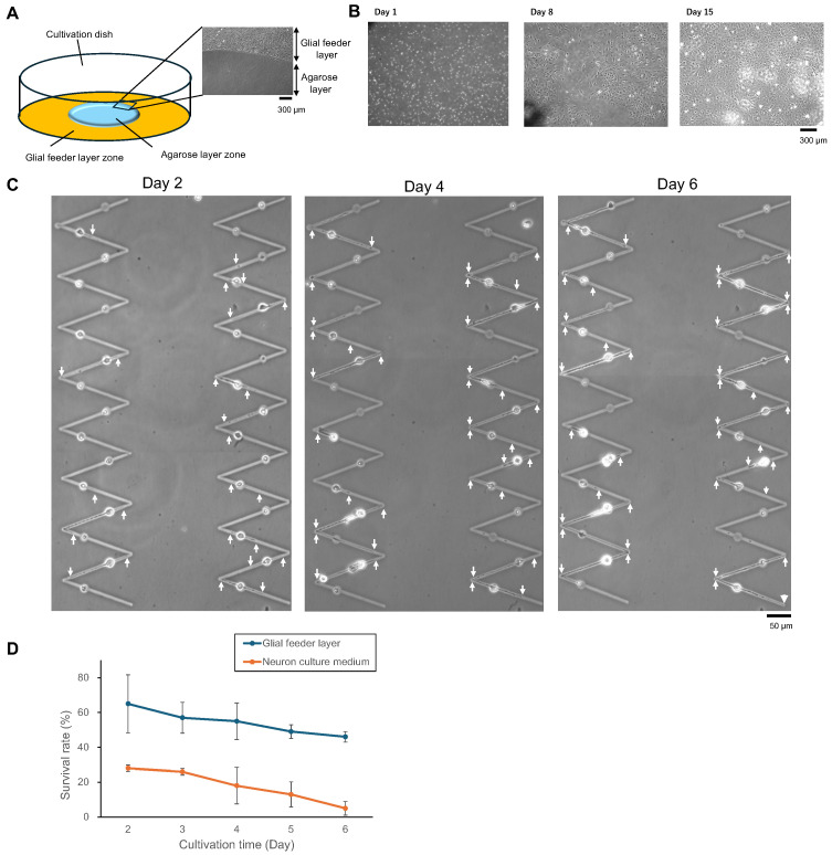
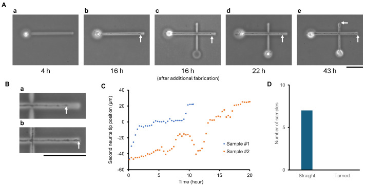
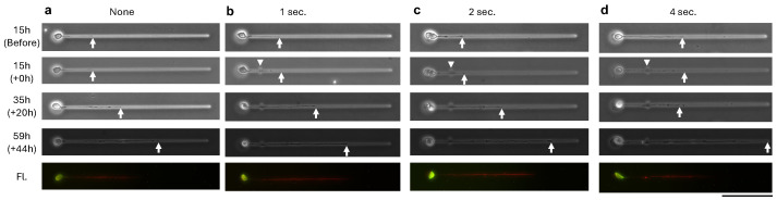
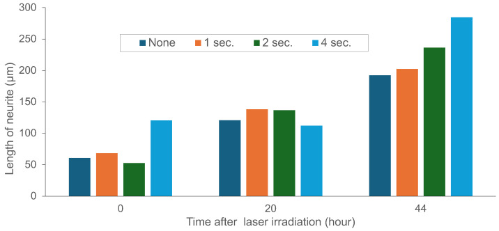
Constructing stable and flexible neuronal networks with multi-neurite wiring is essential for the in vitro modeling of brain function, connectivity, and neuroplasticity. However, most existing neuroengineering platforms rely on static microfabrication techniques, which limit the ability to dynamically control circuit architecture during cultivation. In this study, we developed a modifiable agarose gel-based platform that enables real-time microstructure fabrication using an infrared (IR) laser system under live-cell conditions. This approach allows for the stepwise construction of directional neurite paths, including sequential microchannel formation, cell chamber fabrication, and controlled neurite-neurite crossings. To support long-term neuronal health and network integrity in agarose microstructures, we incorporated direct glial co-culture into the system. A comparative analysis showed that co-culture significantly enhanced neuronal adhesion, neurite outgrowth, and survival over several weeks. The feeder layer configuration provided localized trophic support while maintaining a clear separation between glial and neuronal populations. Dynamic wiring experiments further confirmed the platform's precision and compatibility. Neurites extended through newly fabricated channels and crossed pre-existing neurites without morphological damage, even when laser fabrication occurred after initial outgrowth. Time-lapse imaging showed a temporary growth cone stalling at crossing points, followed by successful elongation in all tested samples. Furthermore, the direct laser irradiation of extending neurites during microstructure modification did not visibly impair neurite elongation, suggesting minimal morphological damage under the applied conditions. However, potential effects on molecular signaling and electrophysiological function remain to be evaluated in future studies. Together, these findings establish a powerful, flexible system for constructive neuroengineering. The platform supports long-term culture, real-time modification, and multidirectional wiring, offering new opportunities for studying neural development, synaptic integration, and regeneration in vitro.

Keywords: agarose gel micropattern; bending angle; bending microchannel array; hippocampal cell; neurite elongation control; neuron; neuronal network formation.


Conflict of interest statement

The authors declare no conflicts of interest.

Figures

Figure 1
Figure 1
Experimental setup and agarose gel microfabrication procedure. (A) Schematic of the custom infrared (IR) laser-based system for agarose gel microstructure fabrication. The setup integrates a fiber-coupled IR laser, live imaging optics, and a motorized X–Y stage for real-time patterning under aqueous conditions. (B) Conceptual diagram of the fabrication sequence: (a) Localized laser melting creates the first cell chamber. (b,c) A microchannel is patterned by translating the stage. (d) A single neuron is manually placed in the chamber. (eg) After several hours of culture, a neurite extends into the microchannel. (hj) A second chamber is formed at the distal end. (k,l) A second neuron is placed, completing a directed two-cell circuit. This configuration enables neurites to cross orthogonally on a two-dimensional plane. (C) Sequential infrared micrographs of the laser fabrication process: (a) Agarose gel-coated dish prior to patterning. (b) Laser irradiation at 0.25 W for 2 s forms the first cell chamber, visualized by a bright laser spot. (c) A circular chamber appears post-irradiation. (d,e) A microchannel is fabricated by moving the stage (0.16 W, 5 μm/s). (fi) Additional microchannels are created using the same process. The visible laser focus facilitates the precise alignment of new features with existing structures. Bright spots of infrared camera images represent the laser spot areas. Scale bar, 100 μm.
Figure 2
Figure 2
Glial co-culture enhances neuronal survival and neurite stability in long-term culture. (A) Experimental setup and representative micrograph showing the two-zone co-culture system. Neurons were seeded on the micropatterns fabricated in an agarose gel-coated zone adjacent to a confluent glial cell feeder layer. The glial cell feeder layer zone surrounds the agarose gel layer zone. The agarose gel layer restricts glial migration, enabling localized neurotrophic support without direct overgrowth. (B) Time-lapse imaging of the glial feeder zone over 15 days. Glial cells reached confluence and maintained a stable monolayer throughout the culture period. (C) Representative time-course images showing neurite elongation and survival of a single neuron co-cultured with glial cells. White arrowheads indicate the tips of elongated neurites, which exhibit progressive elongation from Day 2 to Day 6 after neurons were placed into the agarose microstructures. (D) The time-dependent survival rate of neurons in glial co-culture and neuron culture medium conditions over 6 days after neurons were placed into the agarose microstructures (mean and standard deviation (SD; Error bars), N = 6; N represents the number of cultivation trials). Neuronal survival was significantly enhanced by the presence of glial cells, demonstrating the protective and supportive effects of co-culture.
Figure 3
Figure 3
Stepwise modification of agarose gel microstructures enables sequential multi-neurite wiring and controlled neurite crossing. (A) Time-lapse sequence showing the dynamic fabrication and cultivation of a two-neuron circuit with intersecting neurites. (a) Four hours after cultivation started, the first neuron was placed into a pre-fabricated microchamber. (b) By 16 h, the first neurite fully elongated through the pre-fabricated initial microchannel. (c) After verifying full elongation, a second microchamber and a perpendicular microchannel were fabricated across the path of the first neurite. Then, the second neuron was introduced into the newly formed second chamber. Laser patterning passed directly over the existing first neurite without causing visible damage. (d) At 6 h after the second neuron was seeded, the second neurite initiated extension into the new microchannel. (e) By 43 h, the second neurite successfully crossed the first neurite at the intersection point. White arrows indicate the elongated neurite tips over time. (B) Enlarged time-lapse images of neurite extension following laser-based cross-channel fabrication. (a,b) High-magnification views corresponding to panels (Ad) and (Ae), respectively. (C) Two representative examples of second neurite elongation paths over time. The position of the second neurite tip is plotted relative to the crossing point (set as zero on the Y axis). In both cases, neurites temporarily paused near the intersection before resuming elongation and successfully crossing the first neurite. (D) Summary of neurite crossing behaviors. In all seven tested samples, second neurites successfully crossed the first neurite, confirming the reproducibility of the stepwise wiring strategy. Scale bars: 50 μm.
Figure 4
Figure 4
Time-lapse phase-contrast and fluorescence micrographs of neurite elongation after infrared laser irradiation at neurite contact sites. Cultures were initiated on agarose-patterned substrates, and at 15 h (15 h) post-seeding, laser irradiation was applied directly at the neurite shaft (white arrowhead) for 0 s (a; a non-irradiated control), 1 s (b), 2 s (c), or 4 s (d). Follow-up images were acquired at 35 h (+20 h post-irradiation) and 59 h (+44 h post-irradiation). White arrows indicate neurite tips. The bottom row (Fl.) shows immunofluorescence staining for MAP2 (green) and Tau1 (red) performed at 59 h. Scale bar: 100 μm.
Figure 5
Figure 5
Quantitative analysis of neurite elongation following infrared laser irradiation at neurite contact sites (see Figure 4). Despite exposure durations of up to 4 s, substantially longer than the <1 s pulses used for actual agarose microfabrication (Figure 3), neurites continued to extend without interruption.

Similar articles

References

    1. Hodgkin A.L., Huxley A.F. A quantitative description of membrane current and its application to conduction and excitation in nerve. J. Physiol. 1952;117:500–544. doi: 10.1113/jphysiol.1952.sp004764. - DOI - PMC - PubMed
    1. Bucher D., Goaillard J.M. Beyond faithful conduction: Short-term dynamics, neuromodulation, and long-term regulation of spike propagation in the axon. Prog. Neurobiol. 2011;94:307–346. doi: 10.1016/j.pneurobio.2011.06.001. - DOI - PMC - PubMed
    1. Alcami P., El Hady A. Axonal computations. Front. Cell Neurosci. 2019;13:413. doi: 10.3389/fncel.2019.00413. - DOI - PMC - PubMed
    1. Mateus J.C., Sousa M.M., Burrone J., Aguiar P. Beyond a Transmission Cable—New Technologies to Reveal the Richness in Axonal Electrophysiology. J. Neurosci. 2024;44:e1446232023. doi: 10.1523/JNEUROSCI.1446-23.2023. - DOI - PMC - PubMed
    1. Aebersold M.J., Dermutz H., Forró C., Weydert S., Thompson-Steckel G., Vörös J., Demkó L. “Brains on a chip”: Towards engineered neural networks. Trends Anal. Chem. 2016;78:60–69. doi: 10.1016/j.trac.2016.01.025. - DOI

Grants and funding

LinkOut - more resources