. 2025 Jun 1;11(6):425.
doi: 10.3390/gels11060425.

Gelatin/Cerium-Doped Bioactive Glass Composites for Enhancing Cellular Functions of Human Mesenchymal Stem Cells (hBMSCs)

Affiliations

Gelatin/Cerium-Doped Bioactive Glass Composites for Enhancing Cellular Functions of Human Mesenchymal Stem Cells (hBMSCs)

Andrey Iodchik et al. Gels. .

Abstract

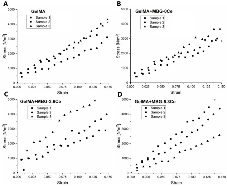
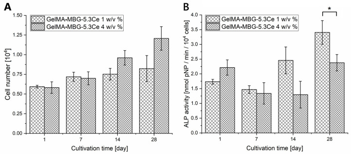
Delayed or non-healing of bone defects in an aging, multi-morbid population is still a medical challenge. Current replacement materials, like autografts, are limited. Thus, artificial substitutes from biodegradable polymers and bioactive glasses (BGs) are promising alternatives. Here, novel cerium-doped mesoporous BG microparticles (Ce-MBGs) with different cerium content were included in photocrosslinkable, methacrylated gelatin (GelMA) for promoting cellular functions of human mesenchymal stem cells (hBMSCs). The composites were studied for intrinsic morphology and Ce-MBGs distribution by scanning electron microscopy (SEM) and energy-dispersive X-ray spectroscopy (EDX). They were gravimetrically analyzed for swelling and stability, compressive modulus via Microsquisher® and bioactivity by Fluitest® calcium assay and inductively coupled plasma-optical emission spectrometry (ICP-OES), also determining silicon and cerium ion release. Finally, seeding, proliferation, and differentiation of hBMSCs was investigated. Ce-MBGs were evenly distributed within composites. The latter displayed a concentration-dependent but cerium-independent decrease in swelling, while mechanical properties were comparable. A MBG type-dependent bioactivity was shown, while an enhanced osteogenic differentiation of hBMSCs was achieved for Ce-MBG-composites and related to different ion release profiles. These findings show their strong potential in promoting bone regeneration. Still, future work is required, e.g., analyzing the expression of osteogenic genes, providing further evidence for the composites' osteogenic effect.

Keywords: cerium-doped mesoporous bioactive glasses; human mesenchymal stem cells; hydrogels; methacrylated gelatin; osteogenic differentiation.


Conflict of interest statement

The authors declare no conflicts of interest. The funders had no role in the design of this study; in the collection, analyses, or interpretation of data; in the writing of the manuscript; or in the decision to publish the results.

Figures

Figure A1
Figure A1
Grouped quartile bar chart representing pore size dispersion for long (red) and short (blue) axes for composites containing 1% MBGs in comparison to pure GelMA. The boxes indicate the first (Q1) and third quartiles (Q3), with the horizontal line inside the box showing the median (Q2) and the small square representing the mean. Whiskers extend to the smallest and largest values within 1.5 times the interquartile range (IQR) from Q1 and Q3. Black diamonds denote outliers beyond this range. Asterisks indicate statistical significance (two-way ANOVA), *** (p < 0.001) and ** (p < 0.01), with horizontal lines showing specific significant comparisons; n = 100 for each composite.
Figure A2
Figure A2
Intrinsic morphology and MBG distribution in composites with different concentrations of Ce-free MBGs. (A) SEM images of GelMA composites containing different concentrations of MBG in comparison to pure GelMA; scale bar: 100 µm. (B,C) Elemental analysis and silicon mapping of the same composites. Si location is superimposed on the images and depicted in cyan color; scale bar: 500 µm.
Figure A3
Figure A3
SEM images of MBG particles. (A,B) MBG-0Ce (scale bars: 200 µm and 30 µm); (C,D) MBG-3.6Ce and (E,F) MBG-5.3Ce (scale bars: 200 µm and 20 µm, both).
Figure A4
Figure A4
Swelling properties and stability of GelMA composites with different concentrations of Ce-free MBGs. (A) Water content and (B) swelling ratio of the composites with different concentrations of MBGs in PBS at 37 °C from 10 to 120 min (n = 4). (C) Mass loss of the hydrogels over 7 days in PBS at 37 °C (n = 4). (D) Images of the freeze-dried composites before (left) and after (right) incubation in PBS for 7 days. Standard deviation is depicted for each bar (n = 4). One-way ANOVA: * p < 0.05, ** p < 0.01, *** p < 0.001.
Figure A5
Figure A5
Stress–strain curves of composites containing 1% w/v of different MBGs. (A) GelMA; (B) GelMA + MBG-0Ce; (C) GelMA + MBG-3.6Ce; (D) GelMA + MBG-5.3Ce. Analysis was performed in comparison to pure GelMA in CCM at 37 °C (n = 3).
Figure A6
Figure A6
Stress–strain curves of composites with different concentrations of Ce-free MBGs. (A) GelMA, (B) GelMA + MBG-0Ce 0.4 w/v %; (C) GelMA + MBG-0Ce 1 w/v %; (D) GelMA + MBG-0Ce 4 w/v %. Analysis was performed in comparison to pure GelMA in PBS at 37 °C (n = 3).
Figure A7
Figure A7
Cell number and specific ALP activity of hBMSCs cultivated on composites containing 1 w/v % or 4 w/v % MBG-5.3Ce; LDH (A) and ALP (B) assays; one-way ANOVA: * p < 0.05; n = 4.
Figure A8
Figure A8
Cell proliferation and osteogenic differentiation. Cell number and specific ALP activity of hBMSCs cultivated in extracts from 1 mg/mL and 4 mg/mL of different MBGs: LDH (A) and ALP (B) assays. n = 2.
Figure A9
Figure A9
Calcium uptake and silicon/cerium release profiles of composites with 1% and 4% MBG-5.3Ce during cell culture of hBMSCs. Accumulated total uptake of calcium (A) and accumulated total release of silicon (B) and cerium (C) were determined by ICP-OES measurements from cell culture supernatants of composites containing 1% w/v compared to 4% w/v of MBG-5.3Ce; n = 4.
Figure A10
Figure A10
Calcium uptake and silicon/cerium release profiles from pure MBGs during cell culture of hBMSCs. Accumulated total uptake of calcium (A) and accumulated total release of silicon (B) and cerium (C) were determined by ICP-OES measurements in extracts from 1 mg/mL of different MBGs; n = 1.
Figure A11
Figure A11
Calcium uptake and silicon/cerium release profiles from pure MBG-5.3Ce during cell culture of hBMSCs. Accumulated total uptake of calcium (A) and accumulated total release of silicon (B) and cerium (C) were determined by ICP-OES measurements in extracts from 1 mg/mL or 4 mg/mL MBG-5.3Ce; n = 1.
Figure 1
Figure 1
Intrinsic morphology and MBG distribution in composites with 1% w/v Ce-free and Ce-MBGs. (A) SEM images of composites (cross-section) containing 1% w/v of MBGs in comparison to pure GelMA. Short and long axis are highlighted with red arrows in the cross-section of GelMA + MBG-5.3Ce composite for illustration. Scale bar: 100 µm. (B) EDX-based silicon mapping and (C) elemental analysis of composites containing 1% w/v of different MBGs. Silicon location is superimposed on the images and depicted in cyan color; scale bar: 250 µm. Characteristic Ce peaks are highlighted with a black arrow.
Figure 2
Figure 2
Swelling and mechanical properties. (A) Water content and (B) swelling ratio of composites containing 1% w/v of different MBGs in comparison to pure GelMA in CCM at 37 °C and 5% CO2 from 10 to 120 min (n = 4). One-way ANOVA: * p < 0.05, ** p < 0.01. (C) Young’s moduli of the same composites incubated at 37 °C and 5% CO2 in CCM (n = 3). (D) Young’s moduli of composites with different concentrations of MBG-0Ce incubated at 37 °C in PBS (n = 3). One-way ANOVA: no statistical differences.
Figure 3
Figure 3
Stability of GelMA composites in CCM. Relative mass increase of composites containing 1% w/v of different MBGs in comparison to pure GelMA without gamma irradiation (left) and gamma-irradiated (right) after 28 days of incubation in CCM at 37 °C and 5% CO2 (n = 3). One-way ANOVA: * p < 0.05. Values between sterilized and non-sterilized gels were compared only for the same material type.
Figure 4
Figure 4
Bioactivity of composites. (A) Calcium content in the destroyed and HCl-treated gamma-irradiated composites containing 1% w/v of different MBGs in comparison to pure GelMA after 28 days of incubation in complete CCM (n = 3). One-way ANOVA: ** p < 0.01, *** p < 0.001. (B) Accumulated calcium uptake of the composites. Differences between cell culture medium before and after incubation with the composites (measured by ICP-OES) was accumulated for every medium change; n = 4. Two-way ANOVA, followed by Tukey’s multiple comparison test, with **** p < 0.0001. (C) SEM images of freeze-dried composites containing 1% w/v of different MBGs in comparison to pure GelMA after 28 days of incubation in CCM; scale bar: 20 µm.
Figure 5
Figure 5
Cell proliferation and osteogenic differentiation. Cell number (LDH assay; (A)) and specific ALP activity (ALP assay; (B)) of hBMSCs cultivated on composites containing 1 w/v % of different MBGs in comparison to pure GelMA. One-way ANOVA: * p < 0.05; ** p < 0.01; *** p < 0.001; n = 4.
Figure 6
Figure 6
Fluorescence microscopic images of hBMSCs on composites after 28 of culture. In the presented Z-stacks, the cytoskeleton is shown in green, and the nuclei in blue. Scale bar: 100 μm. An “s” indicates the direction towards the surface of the composites.
Figure 7
Figure 7
Calcium uptake and silicon/cerium release profiles of composites during cell culture of hBMSCs. Accumulated total uptake of calcium (A) and accumulated total release of silicon (B) and cerium (C) were determined by ICP-OES measurements from cell culture supernatants of composites containing 1% w/v of different MBGs in comparison to pure GelMA; n = 4. Two-way ANOVA, followed by Tukey’s multiple comparison test: * p < 0.05; *** p < 0.001; **** p < 0.0001.

Similar articles

References

    1. Hak D.J., Fitzpatrick D., Bishop J.A., Marsh J.L., Tilp S., Schnettler R., Simpson H., Alt V. Delayed union and nonunions: Epidemiology, clinical issues, and financial aspects. Injury. 2014;45((Suppl. 2)):S3–S7. doi: 10.1016/j.injury.2014.04.002. - DOI - PubMed
    1. Wang C., Shen H., Tian Y., Xie Y., Li A., Ji L., Niu Z., Wu D., Qiu D. Bioactive nanoparticle-gelatin composite scaffold with mechanical performance comparable to cancellous bones. ACS Appl. Mater. Interfaces. 2014;6:13061–13068. doi: 10.1021/am5029582. - DOI - PubMed
    1. Bai X., Gao M., Syed S., Zhuang J., Xu X., Zhang X.Q. Bioactive hydrogels for bone regeneration. Bioact. Mater. 2018;3:401–417. doi: 10.1016/j.bioactmat.2018.05.006. - DOI - PMC - PubMed
    1. Celikkin N., Mastrogiacomo S., Jaroszewicz J., Walboomers X.F., Swieszkowski W. Gelatin methacrylate scaffold for bone tissue engineering: The influence of polymer concentration. J. Biomed. Mater. Res. A. 2018;106:201–209. doi: 10.1002/jbm.a.36226. - DOI - PubMed
    1. Fang X., Xie J., Zhong L., Li J., Rong D., Li X., Ouyang J. Biomimetic gelatin methacrylamide hydrogel scaffolds for bone tissue engineering. J. Mater. Chem. B. 2016;4:1070–1080. doi: 10.1039/C5TB02251G. - DOI - PubMed

LinkOut - more resources