Review
. 2025 Jun 3;11(6):430.
doi: 10.3390/gels11060430.

Multiplexing 3D Natural Scaffolds to Optimize the Repair and Regeneration of Chronic Diabetic Wounds

Affiliations
Review

Multiplexing 3D Natural Scaffolds to Optimize the Repair and Regeneration of Chronic Diabetic Wounds

Cezara-Anca-Denisa Moldovan et al. Gels. .

Erratum in

Abstract

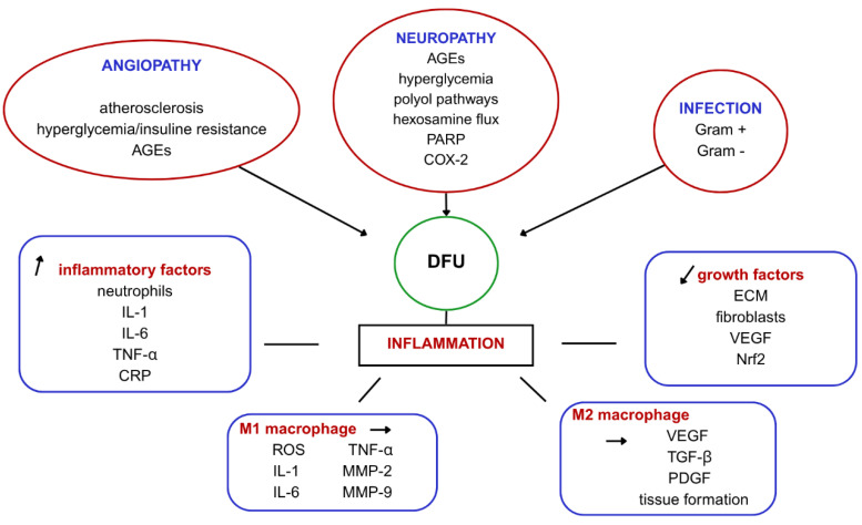
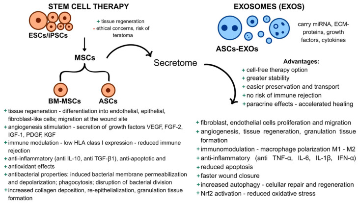
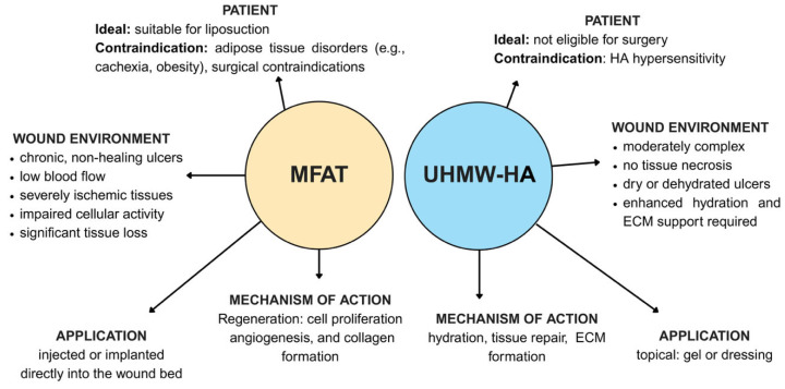
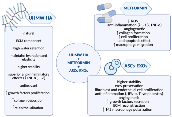
Diabetic foot ulcers (DFU) represent a major complication of diabetes mellitus, affecting millions of patients worldwide and leading to high morbidity and amputation risks. The impaired healing process in DFU is driven by vascular insufficiency, neuropathy, chronic inflammation, and infections. Conventional treatments, including blood sugar control, wound debridement, and standard dressings, have shown limited efficacy in achieving complete healing. Recent advancements have introduced novel therapeutic approaches such as stem cell therapy, exosome-based treatments, and bioengineered scaffolds to accelerate wound healing and tissue regeneration. Mesenchymal stem cells (MSCs), particularly adipose-derived stem cells (ASCs), exhibit anti-inflammatory, pro-angiogenic, and immunomodulatory properties, enhancing wound repair. Additionally, exosomes derived from ASCs have demonstrated the ability to promote fibroblast proliferation, regulate inflammation, and stimulate angiogenesis. The integration of bioengineered scaffolds, including hydrogels, hyaluronic acid (HA), or micro-fragmented adipose tissue (MFAT), offers improved drug delivery mechanisms and a controlled healing environment. These scaffolds have been successfully utilized to deliver stem cells, growth factors, antioxidants, anti-glycation end products, anti-inflammatory and anti-diabetic drugs, or antimicrobial agents, further improving DFU outcomes. This review highlights the potential of combining novel 3D scaffolds with anti-diabetic drugs to enhance DFU treatment, reduce amputation rates, and improve patients' quality of life. While promising, further clinical research is required to validate these emerging therapies and optimize their clinical application.

Keywords: 3D scaffolds; diabetic foot ulcer; drug-delivery; wound healing.


Conflict of interest statement

The authors declare no conflicts of interest.

Figures

Figure 1
Figure 1
The figure illustrates the pathophysiological mechanisms involved in the development and progression of diabetic foot ulcers (DFUs), highlighting key contributing factors such as angiopathy, neuropathy, and infection. Angiopathy, including atherosclerosis and the accumulation of advanced glycation end-products (AGEs), impairs blood flow and tissue healing. Neuropathy, driven by AGEs, hyperglycemia, and the activation of various metabolic pathways, leads to nerve damage and loss of protective sensation. Infection, involving both Gram-positive and Gram-negative bacteria, exacerbates inflammation and delays healing. Inflammation, the central pathological process in DFUs, is driven by pro-inflammatory factors like neutrophils, IL-1, IL-6, TNF-α, and CRP, while M1 macrophages release factors that contribute to tissue damage and chronic inflammation. Impaired healing occurs due to reduced growth factors such as ECM fibroblasts, VEGF, and Nrf2, which delay tissue repair, while M2 macrophages, though promoting healing, are often suppressed in DFUs, further complicating the wound healing process.
Figure 2
Figure 2
Comparison between stem cell therapy and exosome-based therapy in wound healing. On the left, stem cell therapy involves pluripotent stem cells, such as ESCs (embryonic stem cells) and iPSCs (induced pluripotent stem cells), which have high regenerative potential but are associated with ethical concerns and teratoma risks. Stem cells can differentiate into MSCs (mesenchymal stem cells), including BM-MSCs (bone marrow-derived) and ASCs (adipose-derived stem cells), which mediate healing through paracrine signaling (the secretome), releasing growth factors like VEGF, FGF-2, and PDGF to stimulate angiogenesis, tissue regeneration, and immune modulation. MSCs also exhibit anti-inflammatory, anti-apoptotic, and antibacterial effects, enhance collagen deposition, and promote re-epithelialization. On the right, exosome-based therapy uses ASCs-derived exosomes (ASCs-EXOs) as a cell-free alternative. EXOs carry miRNAs, growth factors, and cytokines, offering advantages such as stability, ease of transport, and absence of immune rejection. They accelerate healing by promoting fibroblast and endothelial cell proliferation, angiogenesis, immune modulation, and reducing oxidative stress. EXOs also have anti-inflammatory effects and enhance tissue regeneration and wound closure through Nrf2 activation and autophagy.
Figure 3
Figure 3
Comparative overview of MFAT (micro-fragmented adipose tissue) and UHMW-HA (ultra-high molecular weight hyaluronic acid) as therapeutic options for wound healing. The figure contrasts the ideal patient profiles, wound environments, mechanisms of action, and methods of application for each material. MFAT is best suited for patients who are eligible for liposuction, with contraindications including adipose tissue disorders such as cachexia or obesity, and any surgical contraindications. It is indicated for chronic, non-healing ulcers in ischemic and low-perfusion tissues, when conventional therapies have failed, especially with impaired cellular activity and significant tissue loss. MFAT is applied by direct injection or implantation into the wound bed and promotes regeneration through cell proliferation, angiogenesis, and collagen formation. In contrast, UHMW-HA is appropriate for patients who are not candidates for surgery, with contraindications including hypersensitivity to HA or specific components of the formulation. It is recommended for wounds that are moderately complex, without tissue necrosis, and characterized by dryness or dehydration, where the tissue is not severely ischemic but requires enhanced hydration and ECM support. UHMW-HA is administered topically, either as a gel or dressing, and contributes to wound healing by enhancing hydration, supporting tissue repair, and facilitating ECM formation.
Figure 4
Figure 4
This figure presents a personal approach for a potential future treatment option for diabetic foot ulcers (DFU) using a combination of ultra-high molecular weight hyaluronic acid (UHMW-HA), metformin, and adipose-derived stem cell exosomes (ASCs-EXOs) to enhance wound healing through synergistic mechanisms. UHMW-HA offers superior stability compared to standard HA, promotes hydration, and supports cell migration, collagen deposition, and re-epithelialization, while also reducing inflammation (↓ TNF-α, IL-6) and providing antioxidant effects. Metformin helps reduce reactive oxygen species (ROS), improves insulin sensitivity, stimulates angiogenesis, and promotes collagen formation. ASCs-EXOs contribute to fibroblast and endothelial cell proliferation, immune modulation by promoting M2 macrophage polarization, and extracellular matrix (ECM) reconstruction, shifting the immune response toward tissue regeneration.

Similar articles

Cited by

References

    1. Shaw J., Sicree R., Zimmet P. Global estimates of the prevalence of diabetes for 2010 and 2030. Diabetes Res. Clin. Pract. 2010;87:4–14. doi: 10.1016/j.diabres.2009.10.007. - DOI - PubMed
    1. Armstrong D.G., Tan T.-W., Boulton A.J.M., Bus S.A. Diabetic Foot Ulcers: A Review. JAMA. 2023;330:62. doi: 10.1001/jama.2023.10578. - DOI - PMC - PubMed
    1. McDermott K., Fang M., Boulton A.J., Selvin E., Hicks C.W. Etiology, Epidemiology, and Disparities in the Burden of Diabetic Foot Ulcers. Diabetes Care. 2023;46:209–221. doi: 10.2337/dci22-0043. - DOI - PMC - PubMed
    1. Raja J.M., A Maturana M., Kayali S., Khouzam A., Efeovbokhan N. Diabetic foot ulcer: A comprehensive review of pathophysiology and management modalities. World J. Clin. Cases. 2023;11:1684–1693. doi: 10.12998/wjcc.v11.i8.1684. - DOI - PMC - PubMed
    1. Deng H., Li B., Shen Q., Zhang C., Kuang L., Chen R., Wang S., Ma Z., Li G. Mechanisms of diabetic foot ulceration: A review. J. Diabetes. 2023;15:299–312. doi: 10.1111/1753-0407.13372. - DOI - PMC - PubMed

LinkOut - more resources